|
ENDOCARDIAL
FIBROELASTOSIS |
Endocardial fibroelastosis is characterized by proliferation of both elastic
and collagenous fibers, causing localized or diffuse thickening of the
endocardium (1).
ETIOLOGY |
- Primary (no associated
cardiac anomalies) (2,3). It is the result of a non-structural insult
resulting in thick layers of collagen and elastic fibers in the
endocardium. Can affect either ventricle
(left > right). - Secondary to other cardiac malformations (2).
- Aortic stenosis.
- Coarctation of the aorta.
- Anomalous origin of the left coronary artery from the pulmonary trunk.
- Secondary to metabolic disorders.
- Carnitine deficiency.
- Mucopolysaccharidosis (4).
- Secondary to viral infections.
- Parvovirus (5).
ULTRASOUND |
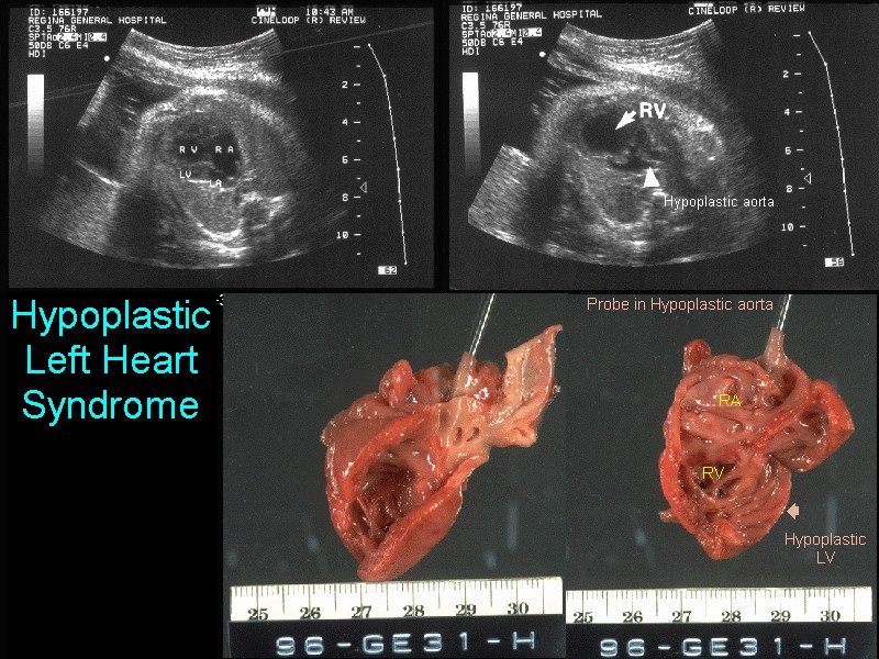
- Usually secondary, associated with left ventricle obstruction, and diagnosed in the second or third trimesters.
- Left ventricle is usually enlarged but may be normal in the less common contracted form.
- 80% present with congestive cardiac failure within the first year of life (6).
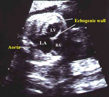
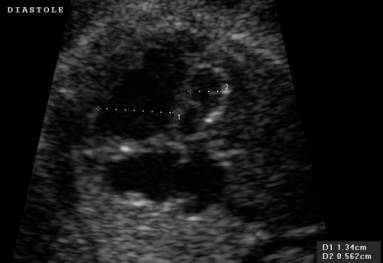
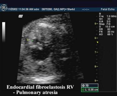
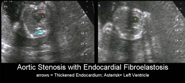
- Thickened, echodense endocardium (2). The hyperechoic rim that forms around the affected chamber is thought to result from blood stagnating within the chamber secondary to outflow tract obstruction. The stagnant blood results in fibrin deposits with the wall of the heart.
- Poor contractility of the ventricle which may lead to fetal hydrops.
- There may be obstruction of the outflow tract by the endocardial thickening (2).
- There may be thickening and narrowing of the ascending aorta and/or the mitral or tricuspid orifice.
|
Endocardial FibroelastosisLeft Ventricle
|
|
Endocardial FibroelastosisLeft Ventricle
|
|
|
|
|
|
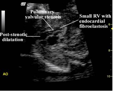
Endocardial Fibroelastosis Right Ventricle
|
|
|
|
|
|
|
|
REFERENCES |
- Moller JH, Lucas RV Jr, Adams P Jr et.al. Endocardial fibroelastosis. A clinical and anatomic study of 47 patients with emphasis on its relationship to mitral insufficiency. Circulation 1964;30:759-782.
- Rustico MA, Benettoni A, Bussani R et.al. Early fetal endocardial fibroelastosis and critical aortic stenosis: a case report. Ultrasound Obstet Gynecol 1995;5:202-205.
- Lurie PR. Endocardial fibroelastosis is not a disease. Am J cardiol 1988;62:468-470.
- Stephan MJ, Stevens EL Jr, Wenstrup RJ et.al. Mucopolysaccharidosis I presenting with endocardial fibroelastosis in infancy. Am J Dis CHILD 1989;143:782-784.
- Anand A, Gray ES, Brown T et.al. Human parvovirus infection in pregnancy with hydrops fetalis. N Engl J Med 1987;316:183-186.
- Wolfson DJ, Pepkovitz SH, van de Velde R et.al. Primary endocardial fibroelastosis associated with hydrops fetalis in a premature infant. Am Heart J 1990;120:708-711.