|
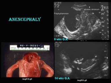
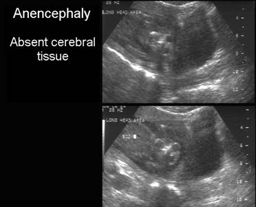
ACRANIA (Absent
Cranial Vault) |
|
|
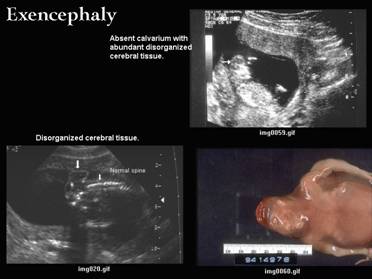
Exencephaly
|
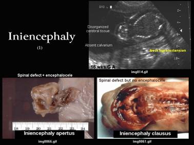
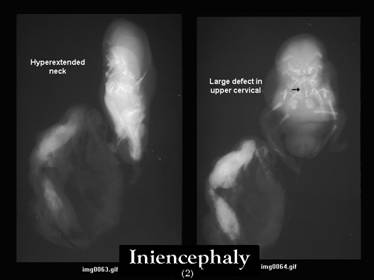
Iniencephaly
|
|
|
Flat bones of calvarium |
Absent |
Partial or Complete absence |
*
Partial or complete absence. Clausus = without encephalocele |
|
|
|
|
|
Skull base
|
N |
N |
*
Dysraphic defect in occiput. |
Fetal neck
|
N |
N |
*
Persistent hyperextension. |
|
Cervical |
N |
N |
*
Hyperextended |
|
|
|
|
|
|
Cerebral tissue |
Absent - occasional remnants of
forebrain |
Present but: |
Normal
or absent if associated with anencephaly or abundant and disorganized if
associated with exencephaly. |
|
|
|
|
|
Brainstem
|
Partially
present esp medulla. |
Present. |
Usually
present. |
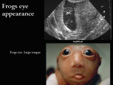
Other signs
|
* Frogs eye: |
|
|
|
|
|
|
|
Pathology
|
Primary
non closure of ant. neuropore or secondary
degeneration of closed neuropore |
?
Failure of mesenchymal migration |
Unknown |
|
REFERENCES |
- Ekici E, Gulmezoglu AM. Sonographic Diagnosis of Fetal Acrania. J Clin Ultrasound 1991, 19:363-366.
- Yang YC, Wu CH, Chang FM et.al. Early Prenatal Diagnosis of Acrania by Transvaginal Ultrasound. J Clin Ultrasound 1992,20:343-345.
- Kennedy KA, Flick KJ,
- Cox GG, Rosenthal SJ, Holsapple JW. Exencephaly: Sonographic Findings and Radiologic-Pathologic Correlation Radiology 1985, 155:755-756.
- Mannes EJ, Crelin ES, Hobbins et.al. Sonographic demonstration of Fetal Acrania AJR 1982, 139:181-182.
- Shere DM, Hearn-Stebbins B, Harvey W et.al. Endovaginal Sonographic Diagnosis os Iniencephaly Apertus and Craniorachischisis at 13 Weeks, Menstrual Age. J Clin Ultrasound 1993, 21:124-127.