|
VOLUME CONTRAST IMAGING (VCI) |
Volume contrast imaging was developed to enhance routine B mode sonography.
The technology is based on real time volume acquisition with surface and maximum gradient surface rendering. The resultant algorithm projects a 3 dimensional data set on a 2 dimensional screen. Because of the small elevation sweep angle, the render box has a large surface area and a relatively small thickness. The user can select the thickness of the render box at 5, 10, 15 or 20 mm.
This rendering process on the slice smoothens the speckle pattern of the image by filling up the gaps with tissue information from the adjacent layer. With surface rendering, pixel from a deeper layer are brought to the surface wherever the gray scale information is not available. This has a smoothing effect on the surface and results in a noise reduction (the proprietary surface algorithm separates and subtracts the noise from the voxel data). Contrast resolution is improved as the noise is reduced. It therefore offers better assessment of:
· Size of a tissue structure.
· Margins of the tissue structure (it enhances the contrast between tissue or organs that would appear similar on conventional 2D ultrasound).
· Internal aspects of the structure.
VCI can be obtained in a number of planes including:
· A – plane – same imaging plane as that obtained at conventional 2D ultrasound.
· C – plane consists of a perpendicular (coronal) plane in relation to the original 2D imaging.
Specific area of study where volume measurements are clinically applicable include:
· Serial fetal lung volume measurements for the prenatal detection of pulmonary hypoplasia.(36-38)
· The volume measurements of the fetal thoraco-lumbar spine as well as kidneys, liver and heart have been established, and fetal liver volumes in normally grown fetuses have been compared to those small-for-gestational-age.(39-44)
· Fetal brain volumes have been calculated using 3D scans with excellent intra and inter observer variability correlating well with other standard biometry at different gestational ages.(45)
· Three dimensional volume measurements may improve our ability to establish fetal weight estimates as well as growth parameters and improve the accuracy with which we can predict small-for-gestational-age infants.
· Fractional limb volume has also been investigated as an additional parameter to further improve accuracy in predicting birth weight, although this remains to be seen in larger scale studies(46-48) Other attempts using 3D to estimate fetal weight have shown superior accuracy due to the inclusion of soft tissue volume measurements, although further work is needed to validate this.(49)
·
Volume measurements of the fetal placenta using
3D sonography has not been particularly helpful to
date in predicting small-for-gestational-age infants.(50)
VOLUME RENDERED IMAGES |
|
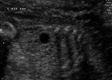
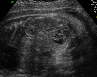
Simple renal cyst |
|
|
|
|
|
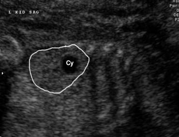
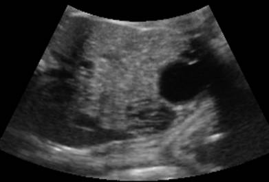
Simple renal cyst – volume
contrast imaging (vci) – c-plane |
|
|
|
|
|
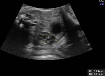
Fetal
ovarian torsion |
|
|
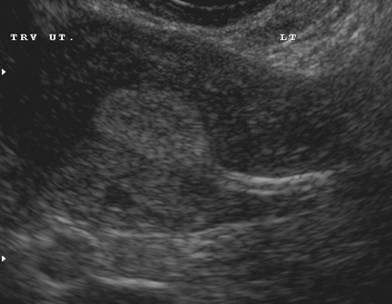
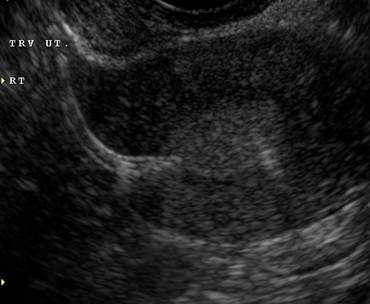
Routine 2D
images |
|
|
|
|
|
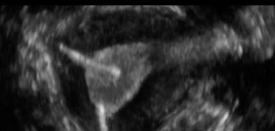
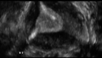
Volume images
(render |
|
|
|
|
|
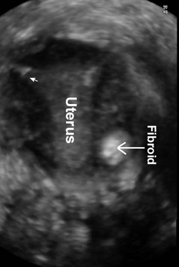
Normal
Uterus |
Fibroid
in myometrium |
|
|
|
|
Essure
sterilization coil insertion |
|
|
Transverse
images through the uterus at the uterotubal
junction |
|
|
|
|
|
Volume
Contrast Image – C Plane |
|
|
|
|
|
|
|


REFERENCES |