|
ARTIFACTS IN 3D ULTRASOUND |
Artifacts arise from a variety of sources in 3D ultrasound. Nelson and co-workers (1) classified them into four broad groups:
- Artifacts due to the 2D ultrasound imaging process.
- Artifacts that arise from the 2D ultrasound process that appear different when viewed from different orientations in the 3D ultrasound volume.
- Artifacts that are unique to 3D ultrasound.
- Artifacts that arise as a result of some operator choice in selecting which part of the volume to display.
These artifacts may arise alone or in combination to produce generated images that may lead to an incorrect diagnosis.
|
2D Artifacts |
|||
|
Type of artifact |
Cause |
2D appearance errors |
|
|
Resolution |
Axial resolution |
Pulse length |
Shape, size |
|
Lateral resolution |
Pulse width |
Shape, size |
|
|
Elevational resolution |
Focusing |
Shape, size (thickened c-plane on 3D) |
|
|
Speckle |
Interference |
Added objects |
|
|
Attenuation |
Shadowing |
Attenuation |
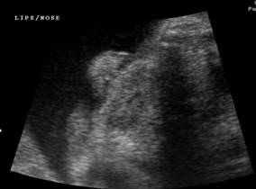
Brightness (3D – pseudocleft; single nostril; nasal bone shadow; partial absence of extremity |
|
|
|
||
|
Enhancement |
Low attenuation |
Brightness (3D -pseudo-narrowing of spine) |
|
|
Refraction |
Refraction |
Brightness |
|
|
Propogation |
Reverberation |
Reflection |
Added objects (3D – multiple intrauterine devices) |
|
Refraction |
Refraction |
Location, shape |
|
|
Mirror image |
Reflection |
Added objects |
|
|
Others |
Comet tail |
Reverberation |
Added objects |
|
Ring - down |
Resonance |
Added objects |
|
|
Speed - error |
Speed - error |
Shape |
|
|
2D color / power artifact |
|||
|
Type of artifact |
Cause |
2D appearance |
3D appearance |
|
Gain |
|
|
|
|
- Flow but no color |
Inadequate gain / sensitivity |
Gap in vessel |
String of pearls |
|
- Color but no flow |
Excessive gain or motion of fluid |
Color in image |
May mimic vessels |
|
- Color bleeding |
Excessive gain |
Large vessels |
Multiple vessels appear as one |
|
- Color noise |
Excessive gain |
Color dots |
Apparent structure |
|
Directional |
|
|
|
|
- Aliasing |
Incorrect velocity range limits |
Misleading color values |
Single vessel may appear multiple |
|
Motion |
|
|
|
|
- Motion flash |
Movement |
Color where no flow |
Streaks / pseudovessels |
|
3D specific imaging artifacts |
|||
|
Acquisition |
|
|
|
|
- Patient movement |
Involuntary movement or respiration |
None |
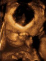
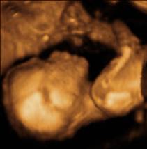
Angled structures e.g. spine or limb. Pseudo cleft |
|
- Organ movement |
Vessel pulsation / cardiac motion |
None |
Pseudo mass; thickened irregular valve or septum; Distorted shapes |
|
Rendering |
|
|
|
|
- Region of interest boundary artifact |
Elimination of important structures |
None |
Hole in skull; absent limb |
|
|
|
|
|
|
- Excessive thresholding artifacts |
Improper setting of thresholds |
None |
Black eyes; micrognathia |
|
- Adjacent structures artifact |
|
None |
Pseudocleft |
|
Editing artifacts |
Elimination of important structures |
None |
Micrognathia; absent limb |
|
|
|
|
|
|
From: Nelson TR, Pretorius
DH, Hull A et.al. Sources and
impact of artifacts on clinical three-dimensional ultrasound imaging. Ultrasound Obstet Gynecol 2000;16:374-383. |
|||

REFERENCES |