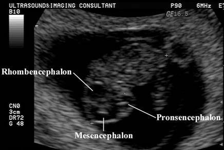
|
ULTRASOUND OF THE
CHOROID PLEXUS |
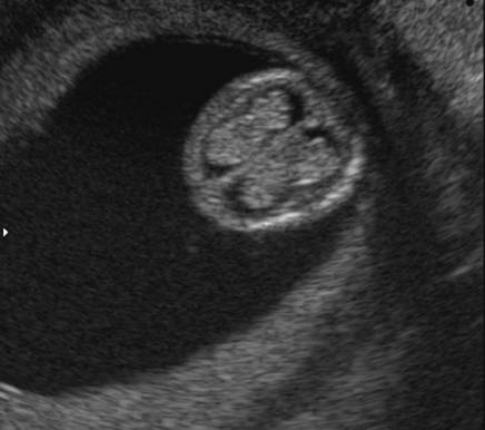
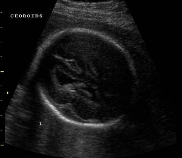
6-7 weeks: Choroid plexus present but not
yet visualized.
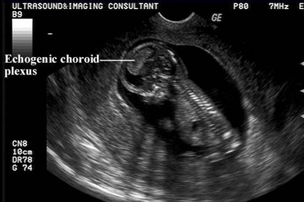
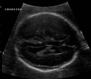
8 weeks: Choroid small and echogenic.
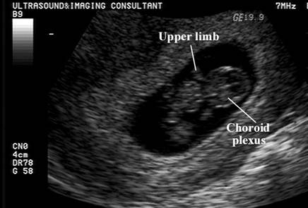
9 weeks: Can be constantly seen on both sides of the falx
within the lateral ventricles. It becomes the most dominant intracranial
structure in the late first trimester cranium.
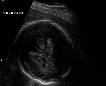
9-11 weeks: Choroid fills the entire lateral
ventricle.
|
|
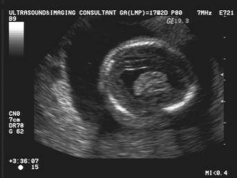
7 weeks |
|
|
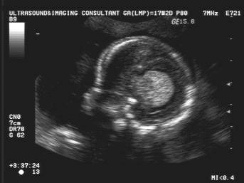
8 wks |
|
|
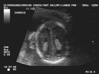
10 wks |
|
|
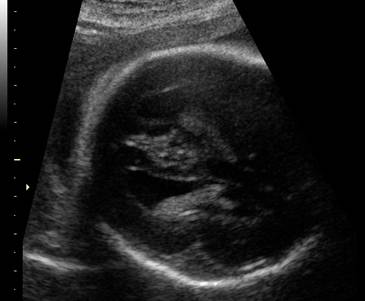
12.5 wks |
12 weeks: Size appears to decrease (due to increased cortical growth of the brain).
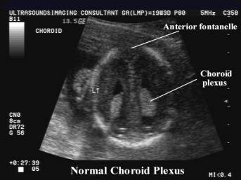
- Best seen in the atria of the lateral ventricles.
- Attached at the Foramen of Munroe.
- Posterior contour is smooth.
- Telia choroidea = thin choroid plexus layer covering the thalami and extending into the lateral ventricles.
- Choroid plexus of the 4th ventricle is difficult to image.
|
NORMAL VARIANTS - DOUBLE CHOROIDAL
PATTERN OR BIFID CHOROID PLEXUS |
The
neonatal choroid plexus may have various sonographic
appearances (1).
A
double choroidal pattern may present as a separation
of the two portions (completely or incompletely), with the medial one
simulating a dangling choroid plexus (2).
The differential diagnosis is an interventricular hemorrhage which usually changes the smooth surface of the choroids.
|
Bifid
choroids plexus |
|
|
|
|
|
|
|
|
NORMAL VARIANTS - THE CALCAR AVIS |
The
calcar avis forms the calcarine
fissure which develops at 16 weeks of gestation.
The
fissure may extend deeply from the medial aspect of the occipital lobe towards
the occipital horn of the lateral ventricles. As the fissure elongates, it folds
and forms a mound of white matter that indents into the medial surface of the
occipital horn, the calcar avis (2,3).
Sometimes
it is more prominent, depending on the depth of the infolding
at the calcarine fissure. In these situations, since
it is isoechoic with surrounding brain tissue, the calcar avis may be confused with a resolving blood clot,
particularly on parasagittal scans. The way to differentiate
it from an intraventricular clot is to slightly tilt
the transducer medially from the cavity of the ventricle. From this view, the calcar avis is properly identified by its continuity with
the brain white matter and branches of the calcarine
fissure.
|
REFERENCES |
- Lee Y, Chung H, Hwang H, Yoon M, Lee H. Choroid plexus in normal full-term neonates: sonographic
classification and clinical application. Presented at the International Pediatric Radiology
Meeting,
- F. F.
Correa, C. Lara, J. Bellver, J et.al. An anatomical fetal brain structure and a normal variant mimicking anomalies on routine neurosonographic imaging: report of two cases. Ultrasound
Obsytet Gynecol
2004;24:672-674
- DiPietro MA, Brody BA, Teele RL.
The calcar avis: demonstration with cranial US. Radiology 1985; 156: 363-364.