|
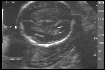
MEASURENT OF
POSTERIOR HORN OF LATERAL VENTRICLE |
- Imaged in an axial plane slightly above the plane used to measure the BPD.
- Area to measure is sonolucent and just posterior to the echogenic choroid plexus.
MEASUREMENTS |
- Posterior Horn Width:
- Measure between echogenic medial and lateral walls.
- Normal = 5-9mm Mean = 7.06mm SD = ± 1.36mm.
|
|
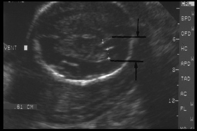
- Cerebroposterior Horn Distance:
- Measure between falx cerebri and lateral walls of posterior horn.
- Increases with advancing gestational age.
- PHW/CPHD ratio decreases with advancing gestational age.
- Important to measure as the posterior horn dilates first and more severely than the rest of the ventricular system.
- Normal Tables (CPHD and PW/CPHD)
|
|
REFERENCES |
- Goldstein I, Reece FA, Pilu G et.al. Sonographic evaluation of the normal developmental anatomy of the fetal cerebral ventricles IV: The posterior horn. Am J Perinatol 1990;7:79-83.