|
Congenital Uterine
Anomalies |
||
|
|
|
|
|
|
|
|
|
|
|
|
|
|
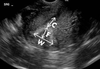
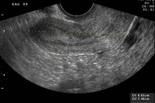
W – measurement of uterine
width F – Fundal distortion C – Length of unaffected
uterine cavity |
Salim R et .al. Reporoducibilty of three
dimensional ultrasound diagnosis Of congemital uterine anomalies. Ultrasound Obstet Gynecol
2003;21:578-582 |
|
Uterine morphology |
Contour of fundus |
External contour |
|
|
Straight or convex |
Convex or Straight Indentation < 10 mm |
|
|
|
|
|
Arcuate |
Concave fundal indentation Central point of indentation at
obtuse angle (>90 0) |
Convex Indentation < 10 mm |
|
|
|
|
|
Subseptate |
Septum which does not extend to
cervix Central point of septum at acute
angle (<900) |
Convex Indentation < 10 mm |
|
|
|
|
|
Septate |
Uterine septum completely
divides cavity from fundus to cervix |
Convex Indentation < 10 mm |
|
|
|
|
|
|
|
|
|
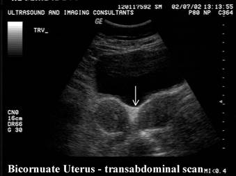
Bicornuate |
Two well formed cornu |
Fundal indentation > 10mm
dividing two cornu |
|
|
|
|
|
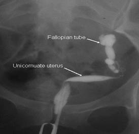
Unicornuate |
Single well formed uterine
cavity with a single interstitial portion of Fallopian tube Concave fundal contour |
Fundal indentation > 10 mm
dividing two cornu if rudimentary horn present |
|
|
|
|
|
|
|
|
|
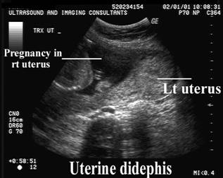
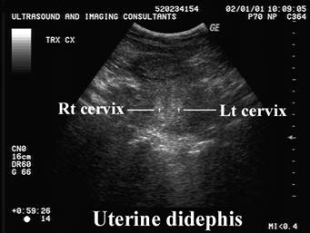
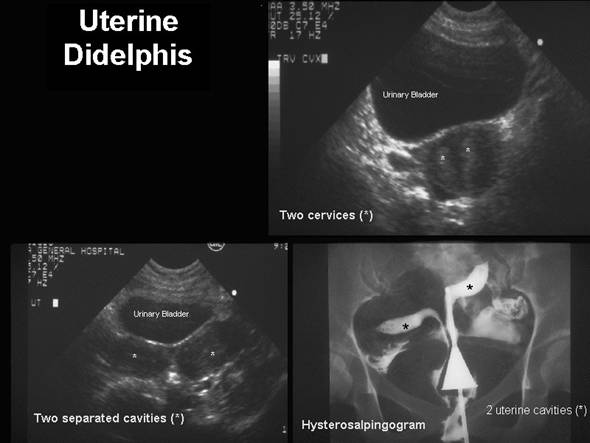
Didelphis |
Two uteri Two cervices Two vaginas |
|
|
|
|
|
|
|
|
|
|
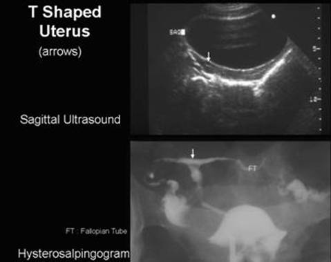
T - shaped |
|
|
|
|
|
|


Acquired
|
|
Link to Myomas
(fibroids) versus Uterine contraction.
|
|
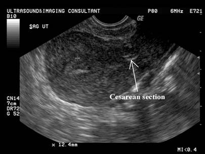
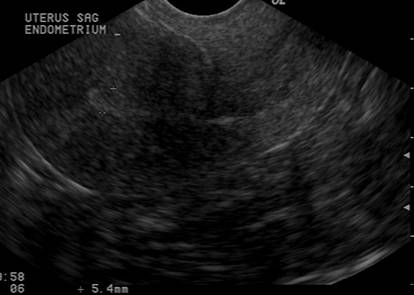
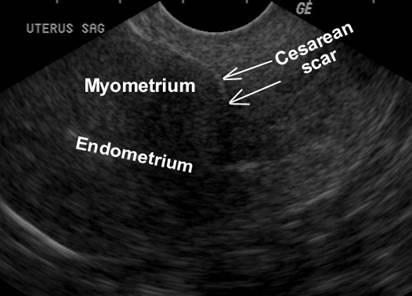
Previous Cesarean Section
|
|
|
|
|
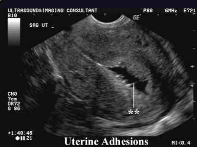
Aschermans
Syndrome.
|
|
Intrauterine
contraceptive device (IUCD) and pregnancy.
|
|
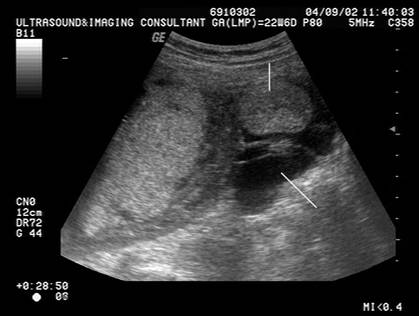
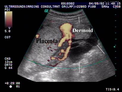
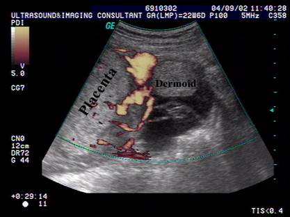
Ovarian
dermoid during pregnancy
|
|