MYOMAS (FIBROIDS) DURING PREGNANCY |
- Incidence in pregnancy is 0.3-2.6% (1).
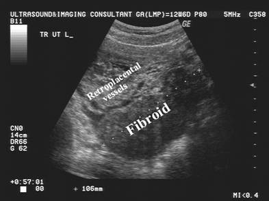
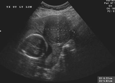
- Solid well-circumscribed mass.
- Hypoechoic.
- May have calcifications.
- May undergo cystic degeneration.
|
|
|
|
|
|
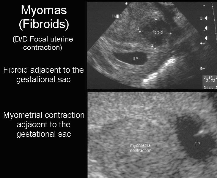
DIFFERENTIAL DIAGNOSIS |
|
Focal Uterine Contraction |
Myoma
|
|
Disappearance after 30-60 min
or on a follow up scan |
No change during the scan or
on a follow up scan |
|
Hypoechoic with respect to myometrium Homogeneous echotexture |
More hypoechoic
than myometrium More heterogeneous echotexture |
|
No attenuation of ultrasound
beam |
Attenuation of ultrasound
beam |
|
May distort endometrial
contour |
Distorts both endometrium and serosal contour
|
|
No calcification Bulges inward |
Calcifications may be present
Bulges both inward and
outward |
|
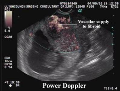
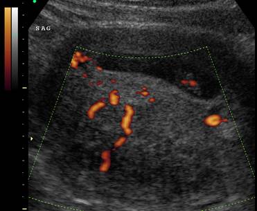
Hypervascular myometrium |
Usually hypovascular
on color doppler (vascular types very rare) |
|
No vessel displacement around
the suspected lesion Blood flow is present
throughout the thickened area of smooth muscle (3) |
Blood vessels splay around
the periphery of the myoma No centralized blood flow is present
(3) within the lesion |
|
|
|
|
|
|
|
|
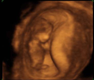
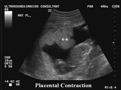
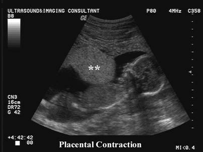
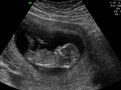
Mild contraction |
|
|
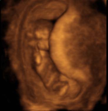
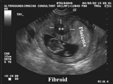
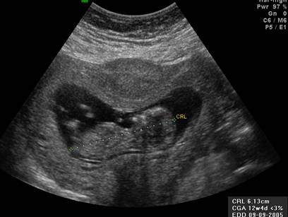
Marked contraction (pressing on the 12 week embryo) |
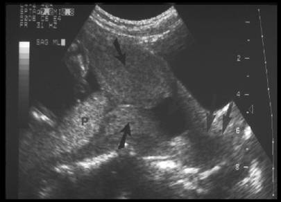
P – Placenta
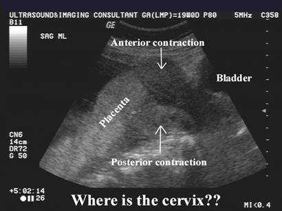
Arrows represent myometrial contractions both
anteriorly and posteriorly Lower arrows delineate the cervix |
Uterine contraction and fibroid in the same patient. Note the change in configuration of the placenta during the scan once
the contraction began resolving |
|
|
|
|
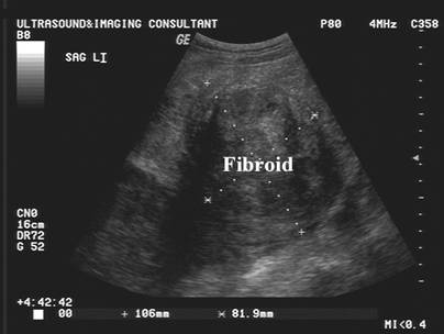
Uterine
contraction and fibroid in the same patient (Case 2) |
|
|
|
|
|
|
|
|
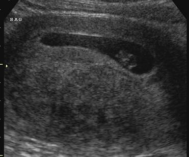
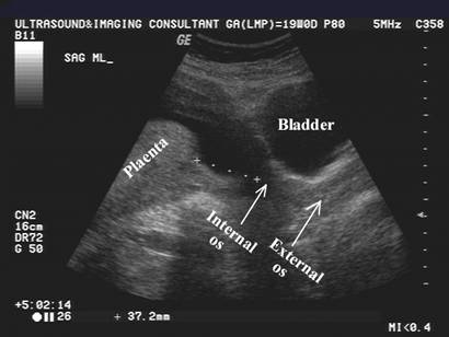
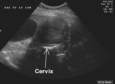
May not be able to delineate the
position of the cervix during a contraction. |
Uterine contraction at 12 wks
4 days which resolved during the scan (image below). |
|
|
|
|
|
|
COMPLICATIONS |
- Increased incidence of spontaneous abortion.
- Increased incidence of premature labor.
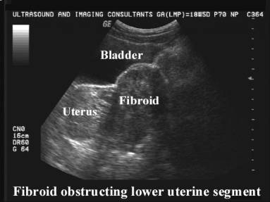
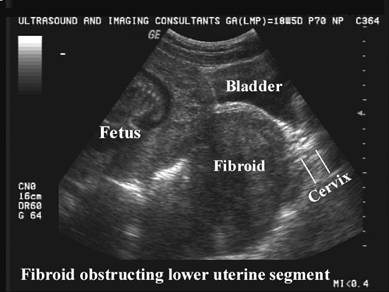
- Obstruction of labor (2) especially if the fibroid is in the lower uterine segment.
|
Case
1 – Lower uterine segment fibroid obstructing the cervix and displacing
the bladder |
|
|
|
|
|
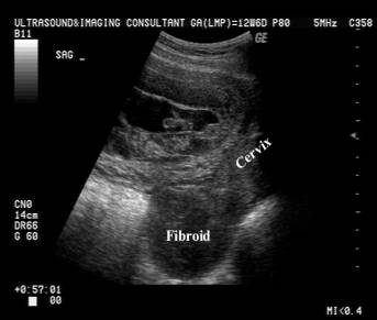
Case
2 – Anterior lower uterine segment fibroid compressing the cervix |
|
|
|
|
REFERENCES |
- Kessler A, Mitchell DG, Kuhlman K et.al. Myoma vs Contraction in Pregnancy: Differentiation with Color Doppler Imaging. J Clin Ultrasound 1993;21:241-244.
- Rice JP, Kay HH . The clinical significance of uterine leiomyomas in pregnancy. Am J Obstet Gynecol 1989;160:1212-1216.
- Trampe BS, Pryde PG, Stewart KS et.al. Color doppler ultrasonography for distinguishing myomas from uterine contractions in pregnancy. J reprod Med 2001;46:791-794.