THE
|
The soft tissues of the fetal neck are easily examined
with transabdominal or endovaginal
ultrasound. The normal appearance is dominated by a single echogenic line, the
dorsal pseudomembrane, first described by Hertzberg
and coworkers in 1989 (1). It is best seen between 10 and 14 weeks gestation
and is thought to represent a spectral reflection of the skin surface along the
back of the fetus simulating a membrane.
ULTRASOUND |
- Dorsal Pseudomembrane - a single pencil thin echogenic line paralleling the occiput and cervical spine. It can be visualized in both a mid sagittal plane or in an axial plane at the level of the cerebellum.
- Anechoic Area - an anechoic
area should present between the pseudomembrane
and occiput. This space should be no more than 3
mm wide when the neck is flexed. Several measurements should be obtained
with the maximum thickness recorded (calipers must be placed on the inner
margins of the lines). After 14 weeks gestational age this anechoic area
becomes progressively more echogenic as subcutaneous tissue and muscle
enlarge.
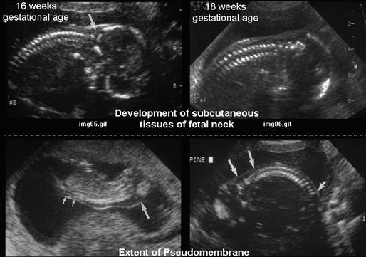
By 16-18 weeks the membrane is no longer seen as a separate entity, but is seen as a nuchal fold contiguous with the underlying soft tissues. - Extent Of Pseudomembrane
- Superiorly - as high as the occiput.
- Inferiorly - usually to L1 or L2, but occasionally to L5.
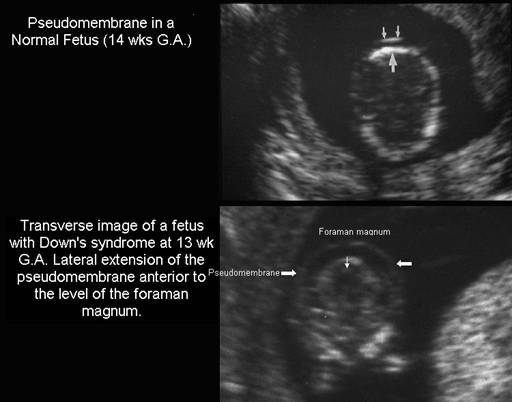
- Laterally - no lateral extension anterior to the posterior rim of the foramen magnum on transverse images.
- Position Of The Fetus - the pseudomembrane can be demonstrated in both the " neck-up " and " neck-down " positions, however, when the membrane is dependent it may be difficult to differentiate from other intrauterine structures, especially the amniotic membrane, or myometrial abnormalities.
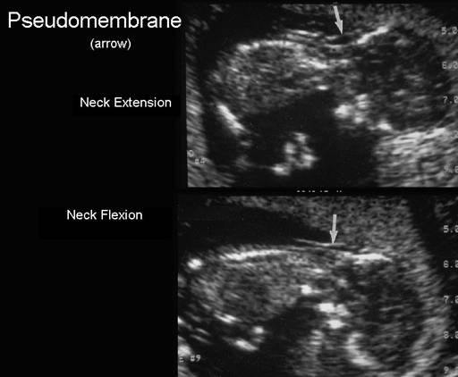
- Neck Flexion - the pseudomembrane is best visualized if the neck is flexed. In the extended position, redundant skin can increase the displacement of the pseudomembrane and simulate a more worrisome nuchal translucency.
- Movement of the membrane - the pseudomembrane moves with the motion of the fetal neck. Other intrauterine membranes such as the normal amnion, amniotic bands and synechiae do not move during fetal neck flexion and extension.
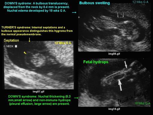
- Septations - septations should be absent.
- Bulbous Swelling - swelling should be absent.
- Fetal Hydrops - hydrops should be absent.
Nuchal translucency refers to the normal subcutaneous fluid-filled space between the back of the fetal neck and the overlying skin. In most cases, this area can be measured accurately and reproducibly on ultrasound between 10 and 14 weeks' gestation.
It is commonly believed that the larger the NT measurement, the greater it's association with Down syndrome, other aneuploidy, major structural malformations, and adverse pregnancy outcome. The etiology of increased NT may be variable, but it is commonly believed to be caused by fluid accumulation in the nuchal region because of aortic isthmic narrowing or other fetal cardiovascular defects, abnormalities in the extracellular matrix, or abnormal or delayed development of the lymphatic system.
|
|
|
Extent
of the pseudomembrane |
|
|
|
Changes
between flexion and extension |
|
|
|
Septations,
bulbous swelling and fetal Hydrops differentiate normality
from pathological conditions |
|
|
REFERENCES |
- Hertzberg BS, Bowie JD,
Carroll BS et al:
- Suchet IB. Ultrasonography of the fetal neck in the first and early second trimesters. Part 1. Normal appearance. Can Assoc Radiol J 1995;46:268-271.