|
NORMAL VALUES FOR THE NUCHAL TRANSLUCENCY AND THE TECHNIQUE FOR MEASUREMENT |
Criteria to maximize good quality of NT ultrasound
- NT ultrasound should only be performed by sinologist / sonographers certified in the technique.
- Transabdominal or transvaginal approach should be left to the sinologist / sonographer's discretion, based on maternal body habitus, gestational age, and fetal position.
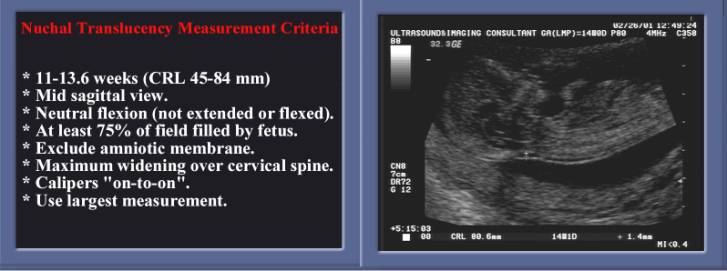
- Gestation should be limited between 10 and 14 weeks (Crown Rump Length (CRL) 36 to 80 mm).
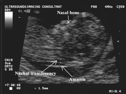
- Fetus should be examined in a mid-sagittal plane.
- Fetal neck should be in a neutral position.
- Fetal image should occupy at least 75% of the viewable screen.
- Fetal movement should be awaited to distinguish between amnion and overlying fetal skin, because, at this gestation, both structures appear as thin membranes. This is achieved by waiting for spontaneous fetal movement away from the amniotic membrane or by manually bouncing the fetus off the amnion.
- Calipers should be placed on the inner borders of the nuchal fold.
- Calipers should be placed perpendicular to the fetal body axis.
- At least three NT measurements should be obtained, with the mean value of those used in risk assessment and patient counseling.
|
|
- Enlarge the image until it nearly fills the screen (>75% of screen).
- Some authors recommend
measuring the nuchal thickness twice and averaging the values (1), while
others advocate measuring it three times and using the largest value for
risk assessment (2). Measure between 11 weeks and 14 weeks. The minimum
fetal crown–rump length should be 45mm and the maximum 84mm. The success
rate for taking a measurement at this gestation is 98–100%, falling
to 90% at 14 weeks; from 14 weeks onwards, the fetal position (vertical)
makes it more difficult to obtain measurements.
- Should be measured in the
neutral position. On the average, the extended nuchal translucency is 0.62
mm greater than the neutral value, while in the flexed position it is on
the average 0.4 mm or less than in the neutral position (3).
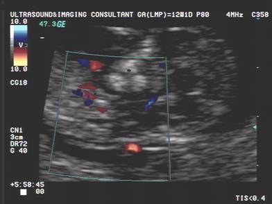
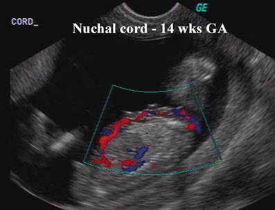
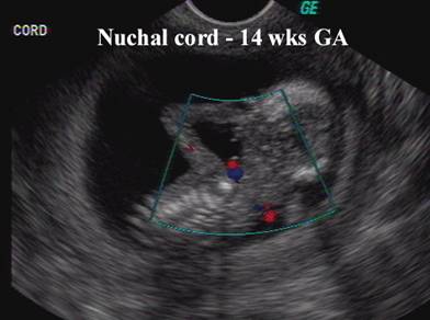
- The umbilical cord may be round the fetal neck in 5–10% of cases and this finding may produce a falsely increased nuchal translucency (may add 0.8mm to the measurement). In such cases, the measurements of nuchal translucency above and below the cord are different and, in the calculation of risk, it is more appropriate to use the smaller measurement.
Nuchal Cord
Note the different thickness of the nuchal translucency
above and below the nuchal cord |
|
|
|
|
|
|
|
|
|
|
- Normal thickness:
- All fetuses develop a measurable nuchal translucency at some point in the first trimester.
- Thickness of the translucency varies with gestational age:
- Peak thickness at 12-13 weeks (in 75% of fetuses).
- At 12-13 weeks the
50th percentile thickness
= 1.7mm. - At 12-13 weeks the
95th percentile thickness
= 2.8mm. - Most authors use a thickness of ³3 mm to define abnormal (some authors use 2.5mm).
- Thickness is independent of maternal age.
- There is a low inter and intra-observer variability. A recent study demonstrated that, after an initial measurement, the second one made by the same (intra-) observer or another (inter-) observer varies from the first by less than 0.54mm and 0.62mm, respectively in 95% of the cases. The study also demonstrated that the caliper placement repeatability was similar to the intra-observer and inter-observer repeatability i.e. a large part of the variation in measurements can be accounted for by the placement of the calipers rather than the generation of the image. Subsequent studies have reported that the intra-observer and inter-observer differences in measurements were less than 0.5mm in 95% of cases.
|
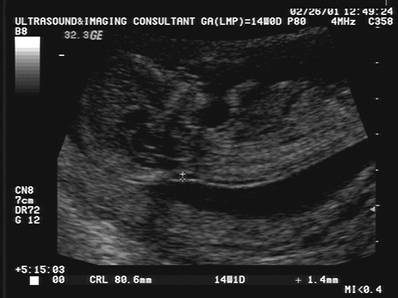
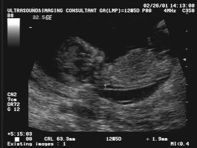
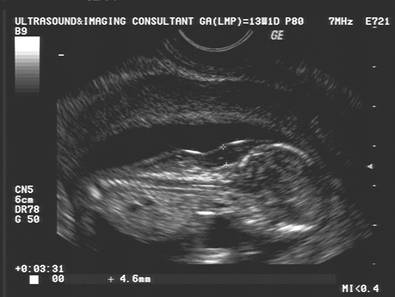
Normal nuchal translucency |
|
|
|
|
|
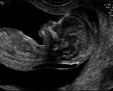
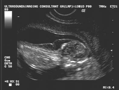
Increased nuchal translucency –
Normal Karyotype |
|
|
|
|
|
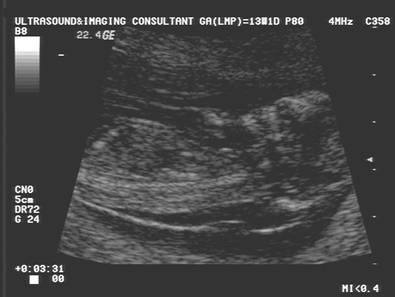
Increased nuchal translucency –
Down Syndrome |
|
|
|
|
|
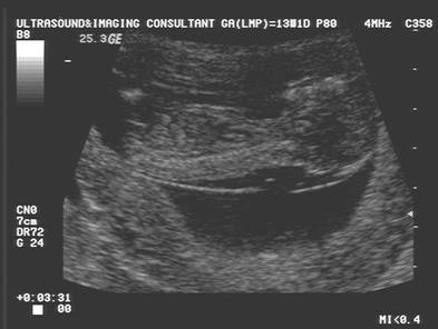
Increased nuchal translucency –
Trisomy 18 |
|
|
|
|
REFERENCES
|
- Panyada PP, Altman D, Brizot ML et.al. Repeatability of measurement of fetal nuchal translucency thickness. Ultrasound Obstet Gynecol 1995;5:337-340.
- Malone FD, D'Alton ME. Fetal nuchal fold translucency screening. Contemporary OB-GYN 1998;43:117-131.
- Whitlow BJ, Chatzipapas I, Economides DL. The effect of fetal neck position on nuchal translucency measurements. Br J Obstet Gynaecol 1998;105:872-876.