|
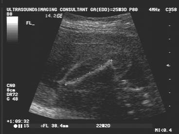
FEMORAL LENGTH |
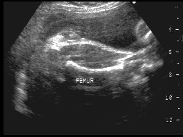
- Its size and ease of measurement makes the femur the long bone of choice in predicting gestational age.
- Transducer should be aligned along the long axis of the bone so that the beam is perpendicular to the shaft.
- Measured ends should be blunt and not pointed.
|
|
|
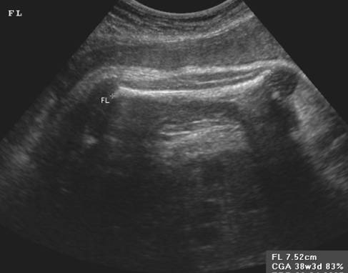
- The length is measured from blunt end to blunt end parallel to the shaft.
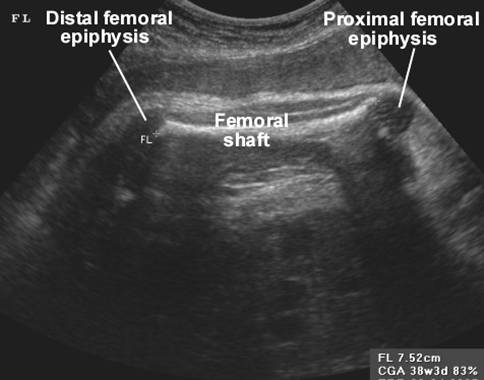
- After 32-34 menstrual weeks the distal femoral epiphysis should be visualized but not included in the measurement.
- Measure the femoral diaphysis, but not the ossification center.
|
|
|
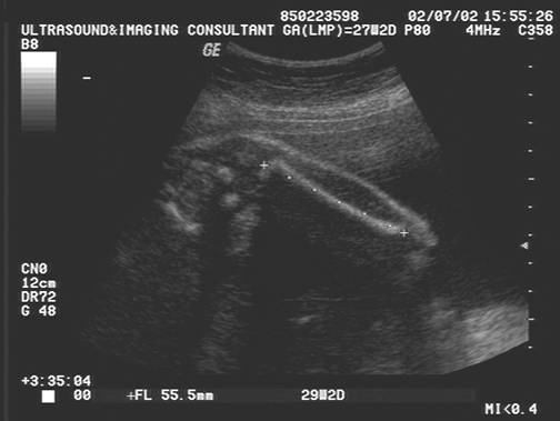
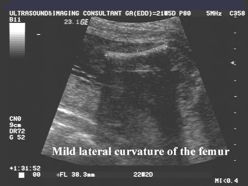
- A minor degree of lateral curvature of the femur is commonly seen in normal fetuses.
|
|
|
- Excessive bowing, demineralization
or shortening excludes the use of the femoral length in assessing
gestational age.
- A normal femur length consistent with expected age without bowing
or angulation is considered adequate for
screening of short limb dysplasias in a patient
with no history of risk factors (1).
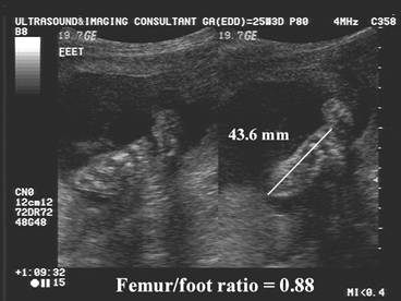
- A femur length to foot length (on
plantar view) ratio > .85 supports familial variation as the cause of a
femur length less than expected (1).
- Variability (+/- 2 SD) (wks).
NORMAL VALUES |
Normal Femur Length
(Table / Graph)
|
Variablilty |
(wks) |
|
12-18 wks |
+/- 1.0 wks |
Precision of predictors of
Gestational Age
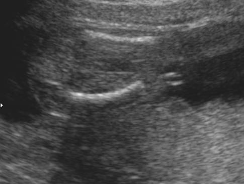
PITFALL |
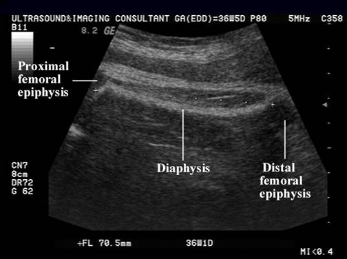
Acoustic shadowing from normal ossification results in the inability to recognize the full thickness of the ossified diaphysis. The cartilaginous ends of the bone help define the true thickness of the diaphysis. The inability to see the full thickness of the diaphysis creates the impression that the femur further from the transducer is bowed (this occurs as only the normally curved medial cortex of the femoral diaphysis is observed).
|
|
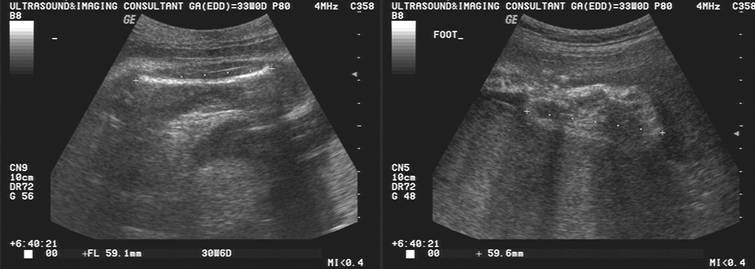
FEMUR / FOOT RATIO |
A normal femur length, consistent with gestational age, and without bowing, angulation or fractures is considered adequate for screening for short limb dysplasia in a patient with no history of risk factors (1).
Normal femur foot ratio of 1.0
|
|
|
|
|
A femur to foot
length (on planter view) ratio of >0.85 supports familial variation as the
cause of a femur less than expected (1).
|
|
|
|
|
REFERENCES |
1.Seeds JW, The Routine or Screening Obstetrical Ultrasound Examination, Clin Obstet Gynecol.
1996; 39(4):824-825.