|
POSTERIOR FOSSA - CISTERNA MAGNA |
- Portion of subarachnoid space that bathes the posterior fossa with CSF.
- Seen as early as 10-14 post menstrual weeks.
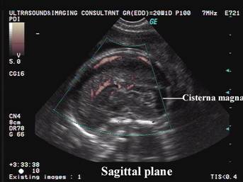
- Echolucent structure posterior to the cerebellum.
- Connection with the IV ventricle through the median aperture can be visualized.
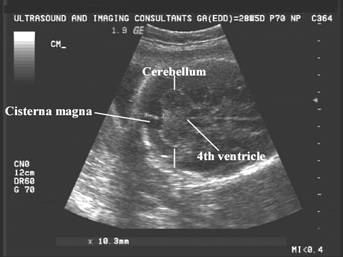
- Arches around cerebellum posteriorly.
- Deepens in midline due to invagination of the vermis.
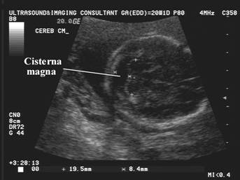
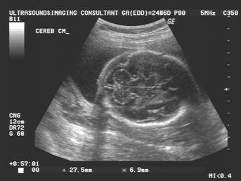
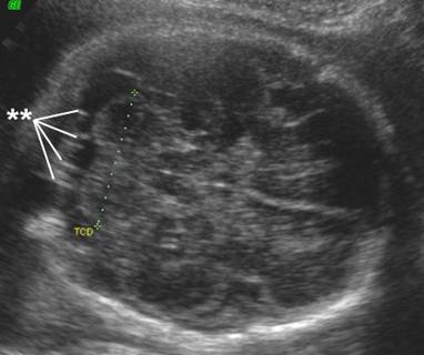
- Measurement (evaluated in the same plane as the cerebellum).
- Measure from posterior aspect of cerebellar vermis to the inner table of the occiput.
- Normal = 5mm SD +/- 3mm (<10mm considered normal) (1-3).
- Constant during pregnancy (Pilu and co-workers) 0.69 ± 0.13cm = 2 SD)
- Linear echoes - may be 1, 2, or many echoes within it (thought to be due to arachnoid).
|
|
|
|
|
|
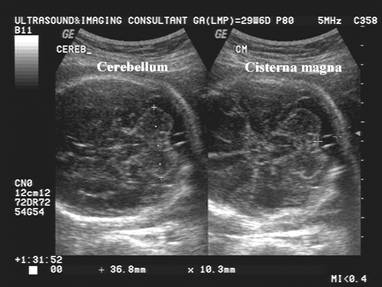
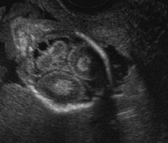
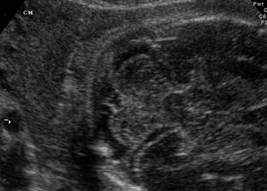
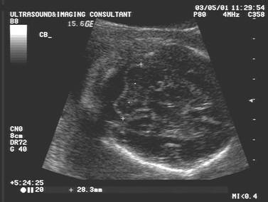
- Giant cisterna magna.
|
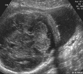
Normal Cisterna Magna |
Giant Cisterna Magna – 13 mm |
|
|
|
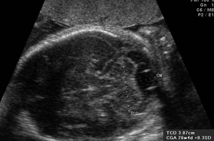
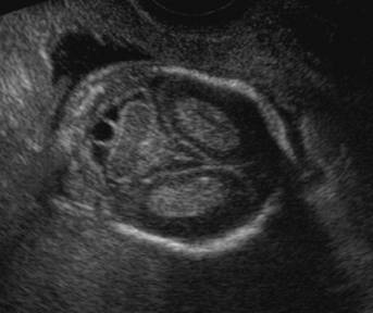
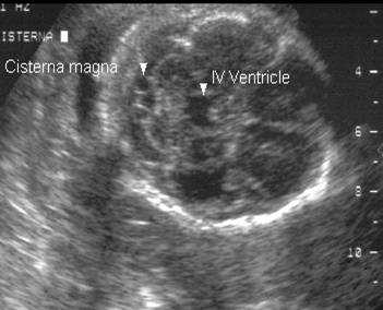
- Pitfall in measurement: Evaluation should be made on an axial image of the posterior fossa which includes the IV ventricle if possible, as angled semicoronal images may falsely enlarge the cisterna magna (2,3).
|
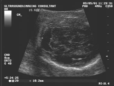
Large Cisterna
Magna (10.2mm)
|
|
|
|
|
REFERENCES |
- Filly RA, Cardoza JD, Goldstein RB et.al. Detection of fetal CNS anomalies: A practical level of effort for a routine sonogram. Radiology 1989;172:403-408.
- Goldstein RB. Sonography of the fetal neural axis: A practical approach. Fetal Maternal Med Rev 1995;7:47-60.
- Laing FC, Frates MC, Brown DL et.al. Sonography of the fetal posterior fossa: False appearance of mega-cisterna magna and Dandy-Walker variant. Radiology 1994;192:247-251.