|
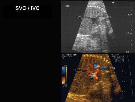
SUPERIOR VENA CAVA
(SVC) INFERIOR VENA CAVA
(IVC) |
- The majority of blood passes through the fetal liver and into the IVC via the hepatic veins.
- 10-50% bypasses the liver and enters the IVC via the ductus venosus.
- 60% of oxygenated blood in the IVC is directed towards the atrial septum via the eustachian valve (which communicates directly with the lower edge of septum secundum). A smaller percentage remains in the RA and mixes with blood from the SVC
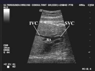
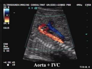
- IVC widens in the proximal portion and enters the RA in a slightly anterior direction. The funnel-like venous confluence is at the end of the abdominal part (venous vestibulum), contains the IVC and orifices of the hepatic veins and ductus venosus.
- It has the appearance of a vertical Y-shaped unit with "2 branches":
- Long branch to LA
- Short branch to RA
- Separated by a cleft between the branches = crista dividens (atrial septum).
- IVC continues as a tube between LA and RA. Walls of this tube are formed by:
- Eustachian valve (also called valve of the IVC) on the right side
- Foramen ovale flap on the left side.
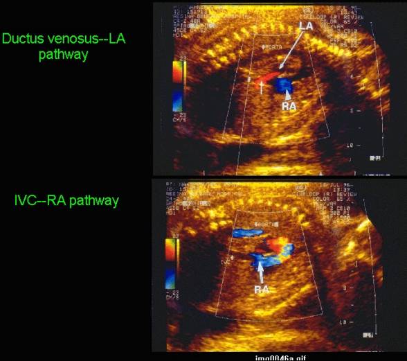
- 2 separate blood pathways result so that the better-oxygenated blood (in the ductus venosus) is directed preferentially towards the brain and coronary arteries.
1. Left sided ductus venosus - foramen ovale pathway that delivers blood directly to the foramen ovale and LA circumventing the RA (oxygenated blood from ductus and left hepatic vein).
2. Right sided IVC - RA pathway - blood is delivered directly to the RA. (poorly saturated blood in IVC enters RA together with SVC blood).
|
|
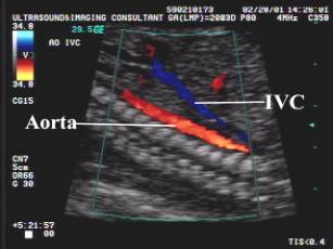
- The left and medial hepatic veins enter the ductus venosus - foramen ovale pathway. The right hepatic veins enter the IVC - RA pathway.
- Inferior Vena Cava Waveform
|
|
|
|
|
|
|
|
|