|
DUCTUS ARTERIOSUS |
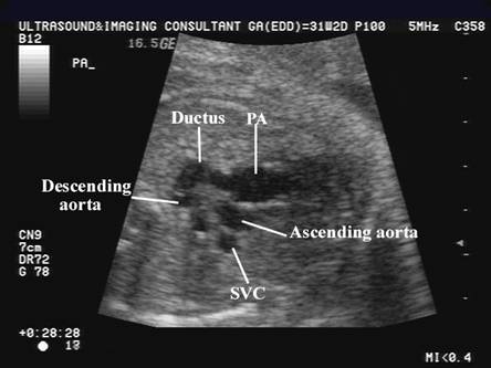
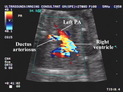
The ductus arteriosus is
a large vessel that connects the pulmonary trunk to the descending aorta in
fetal life. It acts as a right to left shunt at the cardiac level, diverting a
large amount of the combined ventricular output away from the non-functioning
fetal lungs.
- The ductus maintains a short tubular shape, with a caliber that progressively increases with gestation until it's size equals that of the descending aorta at term (10mm) (1).
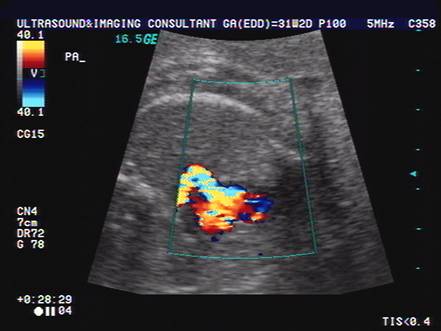
- Ductal peak velocity increases linearly with gestational age and represent the highest velocity in the fetal circulation under normal conditions (2,3).
- Ductus arteriosus flow velocities are the fastest of all the cardiac and extracardiac fetal vessels (2). Blood ejected from the right ventricle into the pulmonary trunk increases in flow velocity as it passes through the ductus (4).
- Normal
Velocities And Pulsatility Index
- Normal Shape.
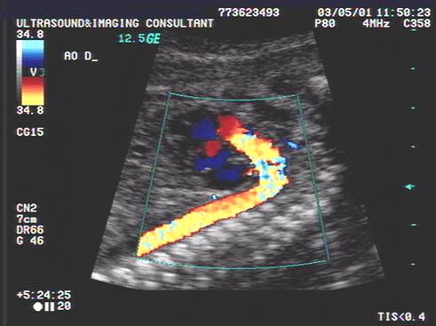
- Straight.
- Mild or sharp curve.
|
|
|
|
|
|
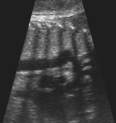
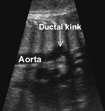
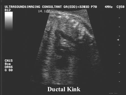
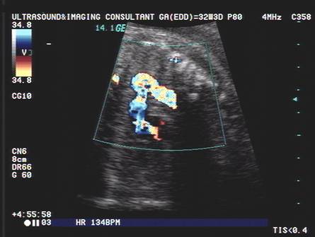
- C or S-shaped (5). Although this has been reported as being caused by tricuspid regurgitation and right heart dilatation (5), other authors (6) point out that the S-shaped trajectory of the ductus is within the normal range of physiological variation (6). This contradiction may be explained by the different planes used by the authors to examine the right cardiac outflow tract (7). The ductus arteriosus can be observed to bend during fetal breathing movements.
|
|
|
|
|
|
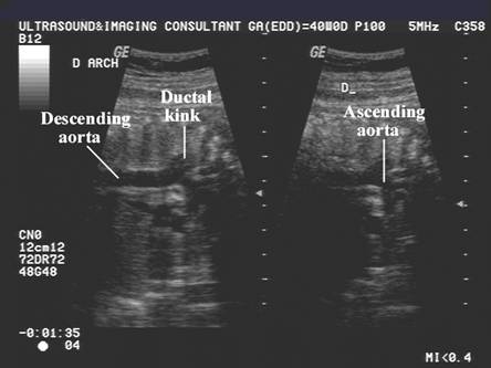
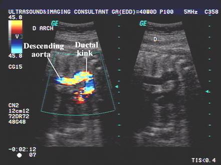
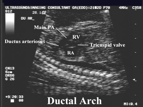
- "Hockey stick" configuration of the ductal arch in a modified longitudinal plane. The arch courses posteriorly and has a "squared off" angle as it enters the descending aorta.
|
|
|
|
|
|
REFERENCES |
- Alvarez L, Aranega A, Saucedo R et.al. Morphometric data on the arterial duct in the human fetal heart. In J Cardiol 1991;31:337-344.
- Huhta JC, Moise KJ, Fisher DJ. Detection and quantification of constriction of the fetal ductus arteriosus by doppler echocardiography. Circulation 1987;75:406-412.
- Van de Mooren K, Barendregt LG, Wladimiroff J. Flow velocity waveforms in the human fetal ductus arteriosus during the normal second trimester of pregnancy. Pediatr Res 1991;30:487-490.
- Brezinka C, Stijnen T, Wladimiroff JW. Relationship between fetal pulmonary trunk and ductus arteriosus flow velocity waveforms in early normal pregnancy. Ultrasound Med Biol 1993;19:527-531.
- Mielke G, Peukert U, Krapp M et.al. Fetal and transient neonatal right heart dilatation with severe tricuspid valve insufficiency in association with abnormally S-shaped kinking of the ductus arteriosus. Ultrasound Obstet Gynecol 1995;5:334-337.
- Benson CB, Brown Dl, Doubilet PM et.al. Increasing curvature of the normal fetal ductus arteriosus with advancing gestational age. Ultrasound Obstet Gynecol 1995;5:95-97.
- Brezinka C. Fetal ductus arteriosus - how far can it bend? Opinion. Ultrasound Obstet Gynecol 1995;6:6-7.