|
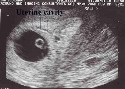
THE UTERUS PRIOR TO
VISUALIZATION OF THE GESTATIONAL
SAC |
IMPLANTATION
OR IMBEDDING OF THE OVUM
|
· Fertilization of the ovum occurs in the lateral or ampullary end of the uterine tube and is immediately followed by segmentation.
· On reaching the cavity of the uterus the segmented ovum adheres like a parasite to the uterine mucous membrane, destroys the epithelium over the area of contact, and excavates for itself a cavity in the mucous membrane in which it becomes imbedded.
· The point of entrance was visible as a small gap closed by a mass of fibrin and leucocytes. The ovum may be covered by a mushroom-shaped mass of fibrin and blood clot.
· All trace of the opening is lost and the ovum is then completely surrounded by the uterine mucous membrane. The structure actively concerned in the process of excavation is the trophoblast of the ovum, which possesses the power of dissolving and absorbing the uterine tissues.
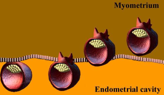
· The trophoblast proliferates rapidly and forms a network of branching processes which cover the entire ovum and invade and destroy the maternal tissues and open into the maternal blood vessels, with the result that the spaces in the trophoblastic network are filled with maternal blood; these spaces communicate freely with one another and become greatly distended and form the intervillous space.
THE DECIDUA
|
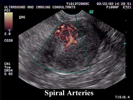
· Before the fertilized ovum reaches the uterus, the mucous membrane of the body of the uterus undergoes important changes and is then known as the decidua. The thickness and vascularity of the mucous membrane are greatly increased; its glands are elongated and open on its free surface by funnel-shaped orifices, while their deeper portions are tortuous and dilated into irregular spaces.
· Decidualized Endometrium.
· The interglandular tissue is also increased in quantity, and is crowded with large round, oval, or polygonal cells, termed decidual cells. These changes are well advanced by the second month of pregnancy, when the mucous membrane consists of the following strata:
o stratum compactum, next the free surface; in this the uterine glands are only slightly expanded, and are lined by columnar cells.
o stratum spongiosum, in which the gland tubes are greatly dilated and very tortuous, and are ultimately separated from one another by only a small amount of interglandular tissue, while their lining cells are flattened or cubical.
o a thin unaltered or boundary layer, next the uterine muscular fibers, containing the deepest parts of the uterine glands, which are not dilated, and are lined with columnar epithelium; it is from this epithelium that the epithelial lining of the uterus is regenerated after pregnancy.
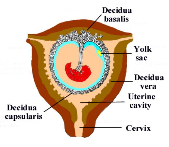
· Distinctive names are applied to different portions of the decidua.
o The part, which covers in the ovum, is named the decidua capsularis.
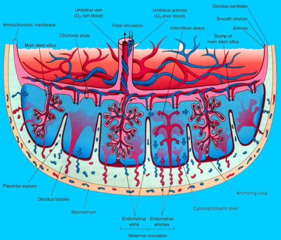
o The portion, which intervenes between the ovum and the uterine wall, is the decidua basalis or decidua placentalis; it is here that the placenta is subsequently developed.
o The part of the decidua, which lines the remainder of the body of the uterus, is known as the decidua vera or decidua parietalis.
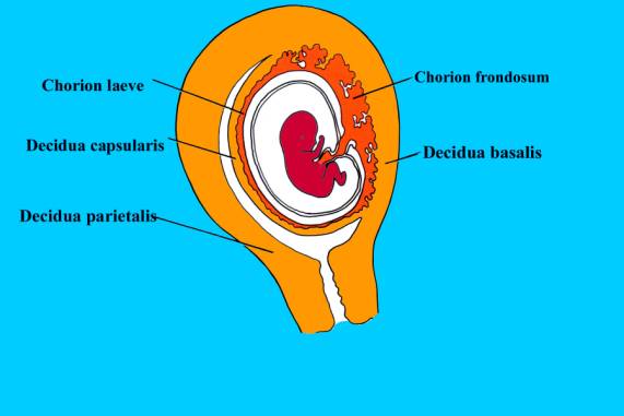
· As the embryo grows, the decidua capsularis is thinned and extended and the space between it and the decidua vera is gradually obliterated, so that by the third month of pregnancy the two are in contact. By the fifth month of pregnancy the decidua capsularis has practically disappeared, while during the succeeding months the decidua vera also undergoes atrophy, owing to the increased pressure. The glands of the stratum compactum are obliterated, and their epithelium is lost. In the stratum spongiosum the glands are compressed and appear as slit-like fissures, while their epithelium undergoes degeneration.
- Decidua basalis (DB) = beneath the implanted embryo.
- Decidua capsularis (DC) = covers the rest of the chorionic sac.
- Decidua parietalis (DP) = endometrial
reaction which lines the uterine cavity and is not involved in
implantation.
- Uterine
cavity (UC).