|
ULTRASOUND ASSESSMENT
OF AMNIOTIC FLUID VOLUME |
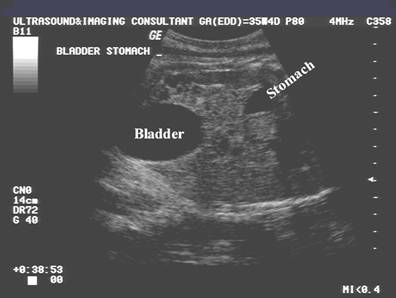
SUBJECTIVE ASSESSMENT |
- The fetus occupies less than half of the intrauterine volume until approximately 22 weeks in the normal pregnancy. Thereafter the fetus progressively occupies a larger proportion of the intrauterine volume.
- This is a qualitative assessment of amniotic fluid volume and is therefore not standardized (1).
- Interobserver and intraobserver variability is reported to be very low.
QUANTITATIVE ASSESSMENT |
- Single Deepest Pocket Measurement (2).
|
|
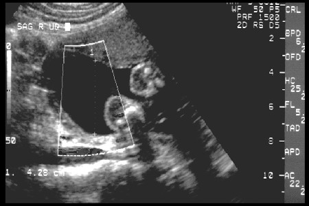
- Measure the dimensions of the largest vertical pocket of amniotic fluid.
- Manning
et.al.1981. : Pocket of fluid
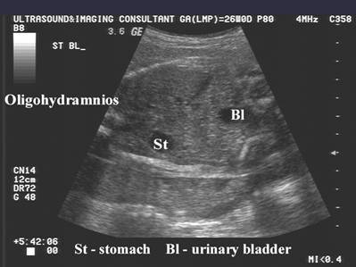
<1cm = oligohydramnios
1-2cm = decreased fluid
2-8cm = normal
>8cm = polyhydramnios (3,4) - Many authors question the 1cm rule as being too restrictive (5).
- Controversies in cut-off criteria for oligohydramnios (6):
- < 0.5 mm (7)
- < 1 cm (8)
- < 2 cm (9)
- < 3 cm (10)
- Amniotic Fluid Index (AFI) (Graphs / Tables) (11-14).
- Technique:
- Divide the uterus into four quadrants using the linea nigra as the vertical axis and the umbilicus as the horizontal axis
- The pocket with the largest vertical dimension is measured in each quadrant.
- Sum of all four measurements = AFI
|
|
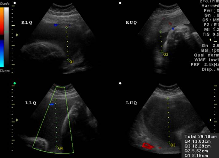
- Values
<5cm = very low (oligohydramnios)
5.1-8cm = low
8.1-25cm = normal
>25cm = polyhydramnios - Controversies in cut-off criteria for oligohydramnios:
- < 5 cm (this represents <1st centile) (12)
- < 5th centile for gestational age (AFI values of 7.1 and 9.7 cm) (15)
- < 7 cm (16)
- < 8 cm (17)
- Others have considered an AFI > 5 and < 10 as borderline. (18).
- Advantages
- Easy to perform.
- More subjective approach than amniotic fluid assessment.
- Requires little training to perform and is ideally suited to real time ultrasound.
- Provides a frame of reference for the inexperienced sonographer.
- Gives a better assessment of amniotic fluid volume than does the single deepest pocket measurement, as the sum of all four quadrants correlate more closely with volume than by using a single measurement.
- Disadvantages
- Wide intraobserver
and interobserver error (20-21).
AFI <5cm - interobserver error = 2cm
AFI >20cm - interobserver error = 5cm - Technical limitations.
- heavy pressure applied by the sonographer with the transducer on the patients abdomen can decrease the height of a pocket of fluid.
- artifacts, especially anterior reverberation artifacts may obscure amniotic fluid situated anteriorly. It may also be difficult to visualize lateral pockets due to the position of the transducer.
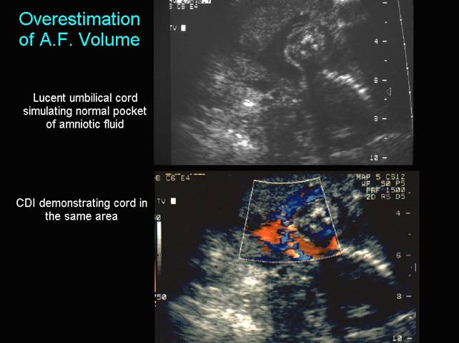
- Overestimation.
- in the third trimester the umbilical cord may be extremely lucent and without duplex or color doppler, cord filled pockets of amniotic fluid may be included in the measurement.
- Dildy et.al. (23) reported that the AFI overestimated the actual fluid volume by as much as 88.7% at lower volumes.
- Chauhan et.al. (24) compared semi-quantitative methods (AFI and two-diameter pocket) with fluid assessment at dye dilution amniocentesis and concluded that the range of the 95% confidence intervals is so wide that sonographic assessment is not a reasonable reflection of the actual amniotic fluid volume.
- Magaan et. al. (25) reported a sensitivity of 6.7% in predicting true oligohydramnios (<500 ml) (25)
- Sonography is imprescise in the detection of oligohydramnios and using multiple assessments did not add to the overall accuracy (26).
|
|
- Fetal movements.
- rapid fetal movements may be a problem as large pockets may be replaced by multiple small pockets between the extremities.
- Pockets with large vertical dimensions and small width's will exaggerate the AFI.
- What is the significance of a full fetal bladder in the presence of ologohydramnios (6). Should this fluid be included in the AFI estimation as it will ultimately be excreted and form part of the fluid estimation.
- Does maternal hydration have any effect on the AFI?
- Two-diameter pocket – vertical X horizontal < 15 cm (22).
REFERENCES |
- Goldstein RB, Filly RA. Sonographic estimation of amniotic fluid volume: Subjective assessment versus pocket measurements. J Ultrasound Med 1988; 7:363.
- Manning FA, Hill LM, Platt LD. Quantitative amniotic fluid volume determination by ultrasound: Antepartum detection of intrauterine growth retardation. Am J Obstet Gynecol 1981; 139: 254-258.
- Chamberlain P. Amniotic fluid volume: Ultrasound assessment and clinical significance. Semin Perinatol 1985; 9:163-167.
- Varma TR, Bateman S, Patel RH et.al. Ultrasound evaluation of amniotic fluid: outcome of pregnancies with severe oligohydramnios. Int J Gynecol Obstet 1988; 27:185-192.
- Hoddick WK, Callen PW, Filly RA et.al. Ultrasonographic determination of qualitative amniotic fluid volume in intrauterine growth retardation. Reassessment of the 1cm rule. Am J Obstet Gynecol 1984; 149:758-762.
- Sherer DM, Langer O. Editorial Oligohydramnios: use and misuse in clinical management. Ultrasound Obstet Gynecol 2001;18:411-419.
- Mercer LJ, Brown LG, Petres RE et.al. A survey of pregnancies complicated by decreased amniotic fluid. Am J Obstet Gynecol 1984;149:355-356.
- Chamberlain PF, Manning FA, Morrison I et.al. Ultrasound evaluation of amniotic fluid volume. I. The relationship of marginal and decreased amniotic fluid volumes to perinatal outcome.Am J Obstet Gynecol 1984;150:245-249.
- Manning FA, Harmon CR, Morrison I et.al. Fetal assessment based on fetal biophysical profile scoring. IV> An analysis of perinatal morbidity and mortality. Am J Obstet Gynecol 1990;162:703-709.
- Halperin ME, Fong KW, Zalev AH et.al. Reliability of amniotic fluid volume estimation from ultrasonograms: intra-observer and interobserver variation before and after the establishment of criteria. Am J Obstet Gynecol 1985;153:264-267.
- Patterson RM, Prihoda TJ, Pouliot MR. Sonographic amniotic fluid measurement and fetal growth retardation: A reappraisal. Am J Obstet Gynecol 1987; 157: 1406-1410.
- Phelan JP, Smith CV, Broussard P, et.al. Amniotic fluid volume assessment with the four-quadrant technique at weeks gestation. J Reprod Med 1987; 32:540-542.
- Rutherford SE, Phelan JP, Smith CV, et.al. The four-quadrant assessment of amniotic fluid volume: an adjunct to antepartum fetal heart rate testing. Obstet Gynecol 1987; 70:353-356.
- Phelan JP,
- Dizon-Townson D, Kennedy
KA,
- Jeng CJ, Lee JF, Wang KG et.al. Decreased amniotic fluid index in term pregnancy: clinical significance. J reprod Med 1992;37:789-792.
- Rodgers MS, Wang CC. A comparison of three ultrasound estimates of intrapartum oligohydramnios for prediction of fetal hypoxia-reperfusion injury. Early Hum Dev 1999;56:117-126.
- Phelan JP, Park YW,
- Rutherford SE, Smith CV, Phelan JP, et.al. Four-quadrant assessment of amniotic fluid volume. J Reprod Med 1987; 32:587-589.
- Banks EH, Miller DA. Perinatal risks associated with borderline amniotic fluid index. Am J Obstet Gynecol 1999;180:1461-1463.
- Chauhan SP, Magaan EF, Morrison JC et.al. Ultrasonographic assessment of amniotic fluid does not reflect actual amniotic fluid volume. Am J Obstet Gynecol 1997;177:291-297.
- Magaan EF, Nolan TE, Hess LW et.al. Measurement of amniotic fluid volume: accuracy of ultrasonography techniques. Am J Obstet Gynecol 1992;167:1533-1537.
- Magaan EF, Chauhan SP,