|
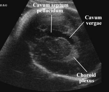
CAVUM SEPTUM
PELLCUDIUM CAVUM VERGAE |
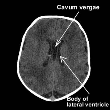
The septa pellucida are two thin transparent leaves that extend from the anterior part of the body, the genu, and the rostrum of the corpus callosum to the superior aspect of the fornix. The cavum septi pellucida (CSP) is a closed cavity in the brain, located on the midline of the transverse plane between the two leaves of the septum pellucidum, which separate the lateral ventricle (1,2). It is thought to arise due to cavitation of the inferior aspect of the commissural plate, presumable around 17 weeks of gestation (3).
Cavum septum pellucidum and its posterior extension cavum vergae are formed simultaneously with the corpus callosum. Visualization is limited to the visualization of the corpus callosum at 17-18 postmenstrual weeks.
- Cavum vergae closes well before term.
- Cavum septum pellucidum usually closes just before term.
|
Incidence of open cavum
septum pellucidum as a function of age (4,5) |
|
|
Age of Fetus (wks) |
% Present |
|
15 wks |
40% |
|
|
|
ULTRASOUND |
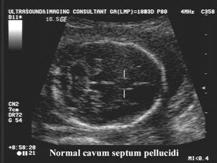
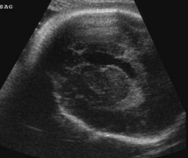
- Median sagittal and mid coronal planes.
- Forms simultaneously with the corpus callosum and is first visualized at 16-18 postmenstrual weeks.
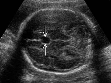
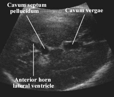
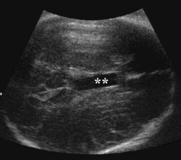
- The cavum septi pellucida is the space between the two leaves of the septum pellucidum whose echogenic walls separate it from the lateral ventricle.
- Cavum vergae – posterolateral extension of the cavum septi pellucida posteriorly between the lateral ventricles.
|
|
|
|
|
|
|
|
|
|
|
CT
Scan |
- Should always be seen when the BPD = 44 mm (4).
- Its absence is associated with midline malformations of the brain (Agenesis of the corpus callosum, holoprosencephaly, septo-optic dysplasia and schizencephaly) (6,7).
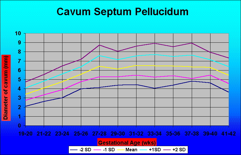
- Cavum
septum pellucidum (Table) – Jou et.al. 1998
- Cavum septum pellucidum (Graph) – Jou et.al. 1998
REFERENCES |
- Johnson ML, Dunne MG, Mack LA et.al. Evaluation of fetal cranial anatomy by static and real time ultrasound. J Clin Ultrasound 1980;8:311-318.
- Thors F, Hoogland HJ. Ultrasonography of the fetal brain: some remarks with respect to the interpretation of the "cavum septi pellucida". J Clin Ultrasound 1990;18:411-414.
- Rakic P, Yakovlev PI.
Development of the corpus callosum and cavum septi in man. J Comp Neurol
1968;132:45-72.
- Falco
P, Gabrielli A, Visentin A et.al. Transabdominal sonography of the cavum
septum pellucidum in normal fetuses in the second and third trimester of
pregnancy. Ultrasound Obstet Gynecol 2000;16:549-553.
- Jou
HJ, Shyu MK, Chen SM et.al. Ultrasound measurements of the fetal cavum
septi pellucidi. Ultrasound Obstet Gynecol 1998;12:419-421.
- Pilu
G, Sandri F, Cerisoli M et.al. Sonographic findings in septo-optic
dysplasia in the fetus and newborn infant. Am J Perinatal 1990;7:337-340.
- Pilu
G, Falco P, Perola A et.al. Differential diagnosis and outcome of fetal
intracranial hypoechoic lesions report of 21 cases. Ultrasound Obstet
Gynecol 1997;9:229-234.