|
THE |
||||||||||||||||||||||||||||||||||||||||||||||||||||||||||||||||||
|
|
||||||||||||||||||||||||||||||||||||||||||||||||||||||||||||||||||
|
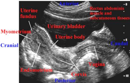
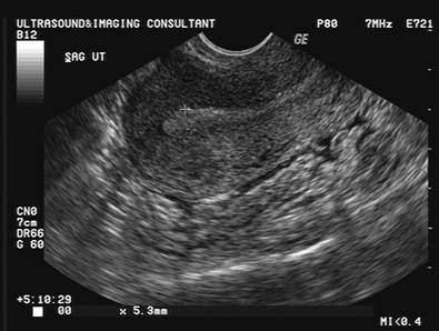
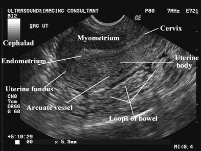
The uterus is a thick, pear-shaped muscular organ located between
the two layers of the broad ligament laterally, the urinary bladder anterior
and the rectosigmoid colon posteriorly.
The myometrium has a
homogeneous echotexture with smooth borders. The
transition between the endometrium and myometrium is delineated by an echo poor line, which is
thought to represent the deeper more vascular layer of the inner myometrium. Normal arcuate
vessels may be seen in the periphery of the uterus. These vessels bifurcate
into radial branches, which supply blood throughout the uterus. |
||||||||||||||||||||||||||||||||||||||||||||||||||||||||||||||||||
|
|
|
|||||||||||||||||||||||||||||||||||||||||||||||||||||||||||||||||
|
|
|
|||||||||||||||||||||||||||||||||||||||||||||||||||||||||||||||||
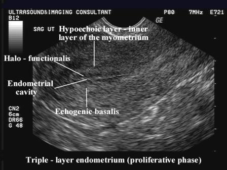
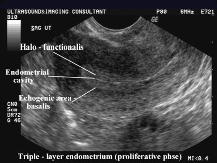
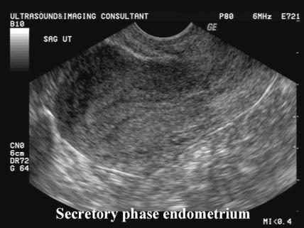
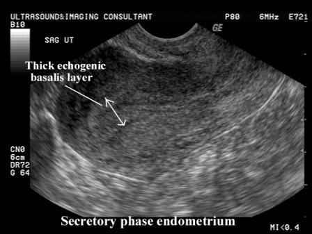
The endometrium appears as a central line whose appearance and density are related to the phases of the menstrual cycle. After menstruation, when the majority of the decidual inner layer of the endometrium has been shed, only a fine cavitatory line present. As the endometrium thickens in the proliferative phase of the cycle, it becomes more prominent but remains echo poor. The endometrium continues to thicken and become more reflective as it enters the secretory phase presumable due to the glands filling with mucin and becoming tortuous. Towards the end of the cycle the endometriual shadow (which includes both the anterior and posterior endometrium) may reach 10 mm. |
||||||||||||||||||||||||||||||||||||||||||||||||||||||||||||||||||
Pre-menarchal
uterus
|
Prior to menarche the uterine body is about
half the length of the cervix. In nulliparas the body and cervix have the same dimensions, while in multiparas the body is approximately double the length of the cervix. |
|||||||||||||||||||||||||||||||||||||||||||||||||||||||||||||||||
|
||||||||||||||||||||||||||||||||||||||||||||||||||||||||||||||||||
Early proliferative
phase
|
|
|||||||||||||||||||||||||||||||||||||||||||||||||||||||||||||||||
Late proliferative
phase
|
|
|||||||||||||||||||||||||||||||||||||||||||||||||||||||||||||||||
Secretory phase
|
|
|||||||||||||||||||||||||||||||||||||||||||||||||||||||||||||||||
|
||||||||||||||||||||||||||||||||||||||||||||||||||||||||||||||||||
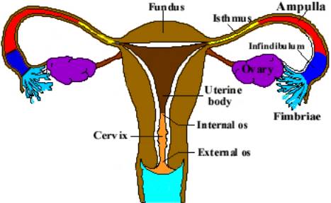
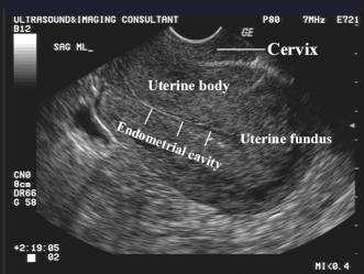
Anatomical segments of the uterus.
- Corpus Uteri (fundus).
- Smooth muscle wall.
- Thickens during pregnancy mainly due to physiologic hypertrophy of the uterine blood vessels. Vessels penetrate all layers of uterus and anastomose freely, emptying into plexuses at the sides of the uterus and broad ligaments.
- Lies between the bladder anteriorly and rectosigmoid colon posteriorly.
- Position is variable and may change with varying degrees of bladder or rectal distention:
- Overfilled bladder tends to result in retroversion.
- Overfilled rectum tends to result in anteflexion.
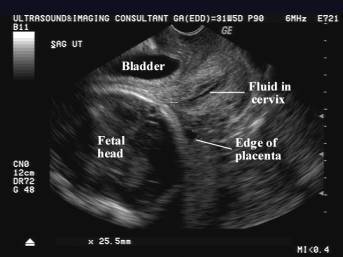
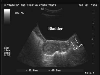
- Cervix Uteri (cervix).
- Posterior to the angle of the urinary bladder.
- This relationship is constant because the cervix is fixed by parametrium at the posterior angle of the bladder.
- Endocervical canal is an echogenic stripe surrounded by hypoechoic fibrous stroma. Length is measured from internal os to its most caudal aspect in the fornix of the vagina.
|
|
|
|
|
|
- Internal os is the fibromuscular junction between the corpus uteri and the cervix. Sonographically they have a similar appearance and cannot be reliable differentiated. It can be localized by its interface with the lower amniotic cavity on longitudinal scans. The amniotic fluid typically is funnel shaped (apex of funnel forms an acute angle at the internal os).
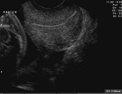
- Normal Length.
- Transabdominal scan = 3.5-5cm. Variation due to compression from a full bladder.
- Transvaginal scan = 4.0-4.5cm.
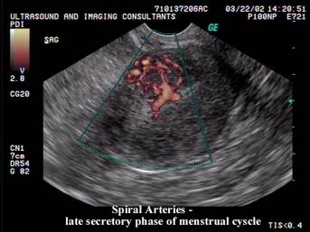
- Uterine vasculature.
- The future uteroplacental arteries, the spiral arteries have a peculiar shape, hence the term "curling arteries" (coined by their discoverer, William Hunter, 1774).
- They are involved with extensive changes during the menstrual cycle they are one of the most labile vascular systems in the human body.
- They arise from the radial arteries at the inner third of the myometrium, while the radials themselves branch from the arcuate system. Small basal arteries arise from the radials and nourish the basal layer of the endometrium, facilitating tissue regeneration after shedding at menstruation or delivery.
- The endometrial segments of the spiral arteries minor sides branches arise, which are the same caliber as the basal arteries, but seem to supply more superficial tissue layers.
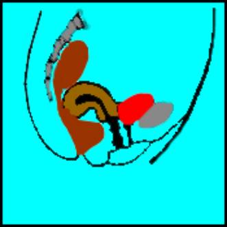
- Terminology for uterine
position:
- Anteversion - cervix courses posteriorly and is directed away from symphysis pubis. Fundus points anteriorly.
- Retroversion – cervix courses anteriorly and is directed away from the symphysis pubis. Fundus points posteriorly.
- Flexion represents the angle between the uterine body and cervix:
- Retroflexion – uterine body posterior at junction of cervix. Almost always associated with retroversion.
- Anteflexion - uterine body curves anteriorly at junction of cervix.
|
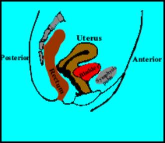
Anteflexion
|
|
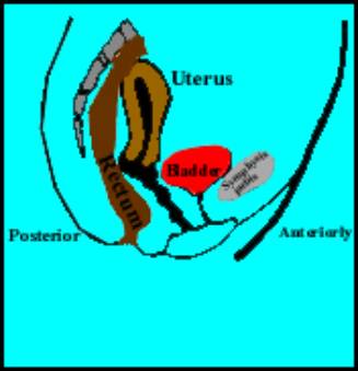
Retroflexion
|
|
|
|
|
Retroversion
+ Retroflexion
|
|
|
|
REFERENCES
|
|
- Harris RD, Barth RA. Sonography of the gravid uterus and placenta: current concepts. AJR 1993;160:455-465.