|
MALE GENITALIA |
|
|
|
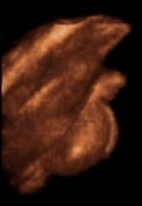
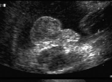
- Parenchyma of mesonephric kidney slowly regresses and the mesonephric tubules make contact with testis cords and rete testis.
- Initial portion of mesonephric duct forms the efferent ductules, while the remaining portion forms the vas deferens. An invagination of the inferior portion for the seminal vesicle.
- Paramesonephric duct regresses by week 8.
- Testis descends within the extraperitoneal mesenchyme along the course of the gubernaculum.
- Normal
Testicular Descent
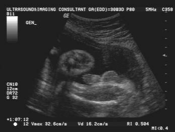
- Bilateral descent was present in 91% of cases, whereas unilateral descent was present in 7% of fetuses by 35 weeks (1).

- External genitalia are sonographically similar until about week 12 of gestation (two pairs of genital folds).
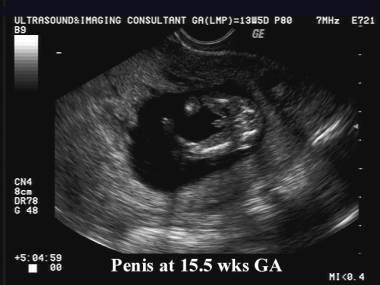
- Medial pairs of folds elongate and fuse along the midline enclosing the urethra.
- Lateral folds enlarge, forming the scrotum.
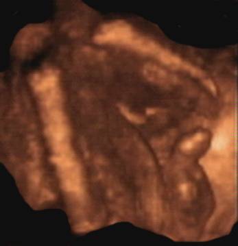
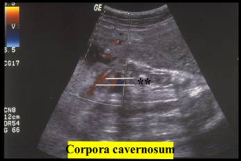
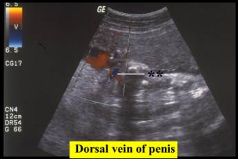
- The urethra may be seen as an echogenic line in the erect penis.
|
|
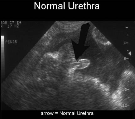
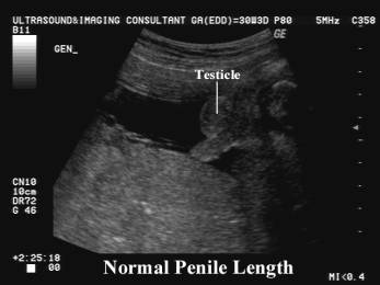
- Penile length has been measured and may be important in cases of bladder outlet obstruction to assist in the differentiation between urethral agenesis and posterior urethral valves (2,3).
- Mean penile length at 14 weeks of gestation is
3.88 mm (90
 CI 2.63-5.39) and reaches up to 23.77 mm (90
 CI 20.51-27.33) at 38 weeks.
|
|
|
|
|
|
|
|
|

REFERENCES |
- Simindokt Z, Gerrard PS, Wise G. Sonographic assessment of fetal male genitalia. JDMS 1991;7:205-207.
- Johnson P, Maxwell D. Fetal penile length. Ultrasound Obstet Gynecol 2000;15:308-310.
- Zalel Y, Pinhas-Hamiel S, Lipitz S et.al. The development of the fetal penis – an in utero sonographic evaluation. Ultrasound Obstet Gynecol 2001;17:129-131.