|
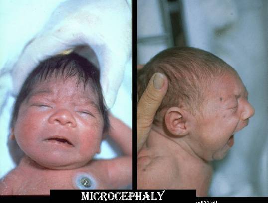
MICROCEPHALY |
Small head, and is usually a product of a small underdeveloped brain, when compared
to the age and body size of the fetus.
|
Genetic Causes |
Environmental |
|
|
|
ULTRASOUND |
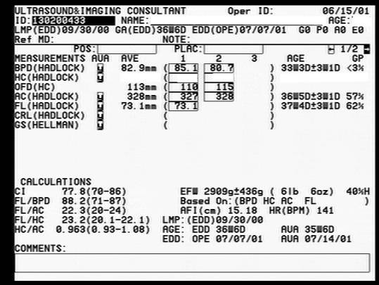
- A small fetal head circumference (below 5th centile corrected for age sex and race). Problem: 5% of normal infants with a small head may be considered microcephalic.
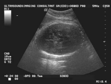
- Tables and Graphs of Biparietal Diameter. >3 SD below the mean for age and sex of the fetus.
- Table and Graph of Ocipitofrontal diameter < 4SD of the predicted mean (2).
- Tables and Graphs of Head circumference (perimeter) was < 5SD of the predicted mean (2).
- Frontal lobe measurements. Anatomic shortening of the frontal lobe appears to precede microcephaly. The frontal lobe is the area of the brain that is most affected and results in the high-sloped forehead. Transcerebellar diameter / Frontal lobe of brain * > 90th centile in some fetuses with microcephaly (3).
- Frontal lobe of brain measurement = linear measurement from back of cavum septum pellucidum to inner table of calvarium midline.
- Deviation of normal blood flow distribution to the fetal brain, especially the frontal lobe in microcephaly. Blood flow in the anterior cerebral arteries is either decreased or not visualized (4)
- Large subarachnoid spaces overlying cerebral convexities, especially the frontal area (5).
- Shape of the lateral ventricle may appear more rudimentary than is expected for that gestational age (absent occipital horns and large ventricular atrial measurement, an appearance that is normally only present prior to 16 weeks gestation) (6).
- Other criteria include:
- Small head circumference.
- Abnormal head/ abdomen circumference ratio.
- Small femur/head circumference ratio.
- Small frontal lobe size. In some cases of microcephaly there is underdevelopment of the forebrain especially the frontal lobes when compared to the rest of the brain or cerebellum (growth spurt in the cerebellum occurs later than that of the forebrain).
|
Head circumference - 33
wks 3 days |
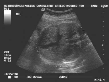
Abdominal circumference
– 36 wks 4 days |
|
|
|
|
|
|
|
|
|
|
REFERENCES |
- Bromley B, Benacerraf B. Difficulties in the Prenatal Diagnosis of Microcephaly. J Ultrasound Med 1995, 14:303-305.
- Chervenak FA, Rosenberg J, Brightman F et.al. A prospective study of the accuracy of ultrasound in predicting fetal microcephaly. Obstet Gynecol 1987;69:908-910.
- Persutte WH, Coury A, Hobbins JC. Correlation of fetal frontal lobe and transcerebellar diameter measurements: the utility of a new prenatal sonographic technique. Ultrasound Obstet Gynecol 1997;10:94-97.
- Pilu G, Falco P, Perolo A et.al. Prenatal diagnosis of microcephaly assisted by vaginal sonography and power doppler. Ultrasound Obstet Gynecol 1998;11:357-360.
- Libicher M, Troger J. Us measurements of the subarachnoid space in infants: normal values. Radiology 1992;184:749-751.
- Goldstein I, Reece EA, Pilu G et.al. Sonographic evaluation of the normal developmental anatomy of the fetal cerebral ventricles. IV. The posterior horn. Am J Perinatol 1990;7:79-83.