|
FETAL AZYGOUS
VEIN |
EMBRYOLOGY |
The azygous systems of veins are derived from the embryonic supracardinal system. The system is paired, and develops during the 6-7th week of gestation, and runs parallel to the paravertebral sympathetic chain:
- The left supracardinal vein becomes the hemiazygous vein.
- The right supracardinal vein gives rise to the entire azygous vein except its cephalic terminus (derived from the distal portion of the posterior cardinal vein). This joins the anterior cardinal vein (the embryologic SVC) forming the azygous arch.
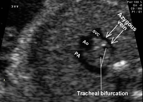
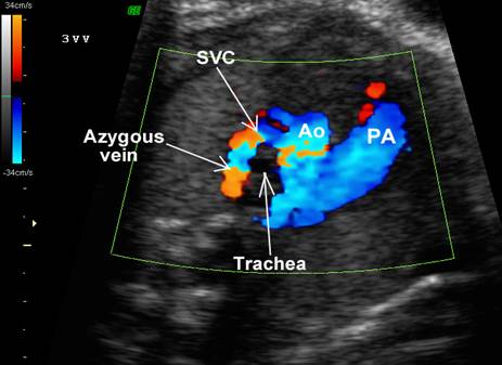
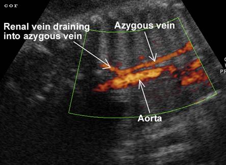
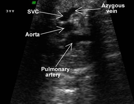
ANATOMY |
The thoracic azygous vein arises from the right
ascending lumbar vein and passes into the thorax via the aortic hiatus. It
always travels to the right of the vertebral column, and arches anteriorly over the root of the right lung superiorly to
end in the SVC. The azygous vein receives all its
blood from all but the first of the right intercostal
veins, the esophageal veins, pericardial veins, right bronchial and subcostal veins.
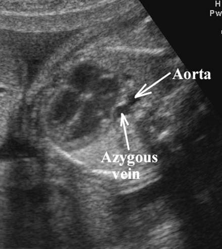
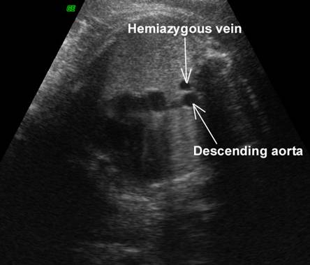
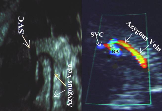
- Anechoic fluid-filled vascular linear channel:
- Anterior to the vertebral column.
- To the right of the fetal thoracic aorta.
- Arches anteriorly to join the SVC.
|
|
|
|
|
|
- Size.
- 1-2 mm up to 30 weeks gestation (seen in 50% of cases).
- 2-4 mm during the last 10 weeks of pregnancy (seen in 98% of cases).
- Enlarged in fetal azygous continuation of the IVC.
|
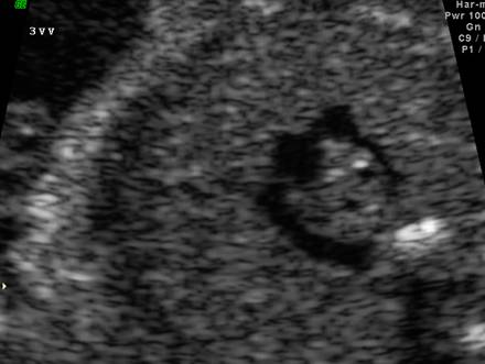
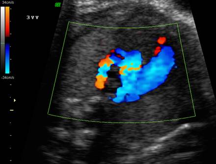
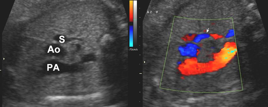
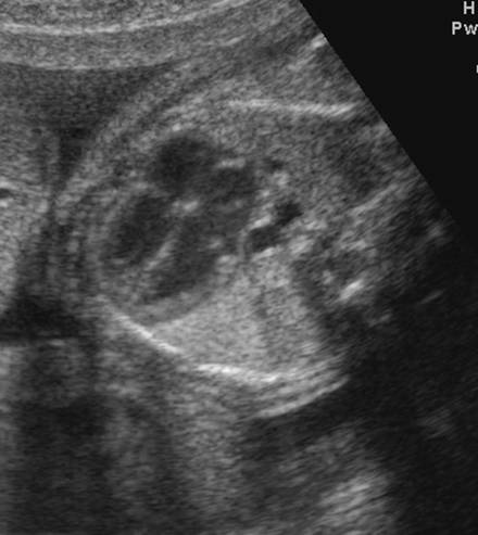
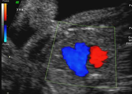
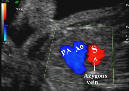
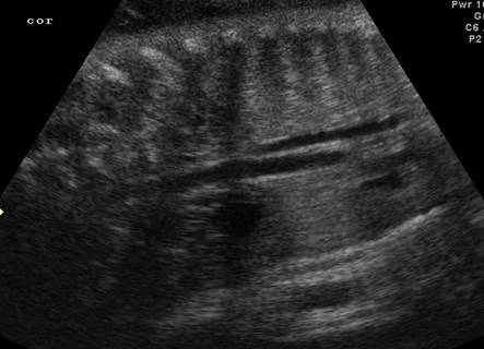
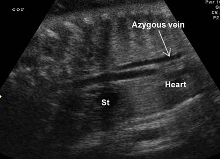
Normal azygous
vein at level of the three vessel view (upper mediastinum) |
|
|
|
|
|
|
|
|
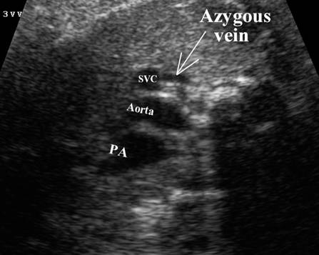
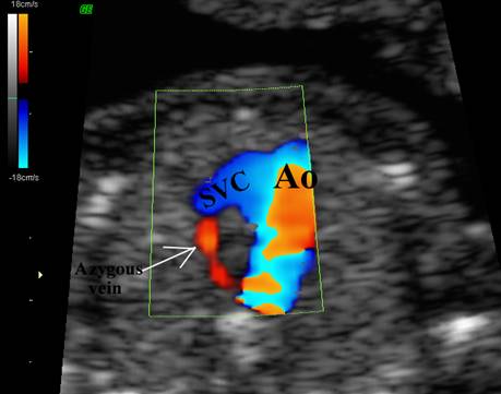
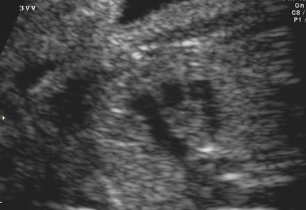
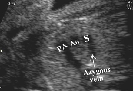
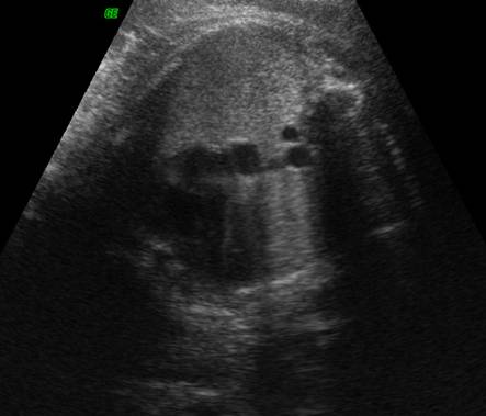
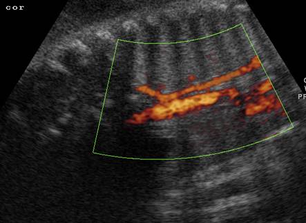
Sagittal views of the normal azygous vein |
|
|
|
|
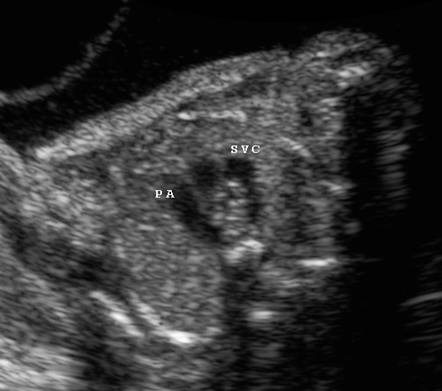
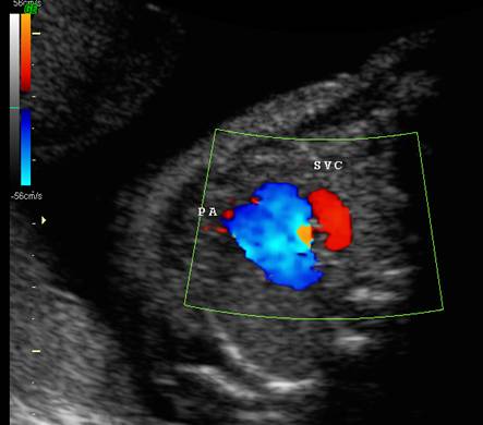
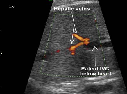
AN
Congenital IVC
obstruction with azygous continuation
Note the two vessels behind the fetal
heart and the large azygous vein draining into the SVC on the three vessel view. |
|
|
|
|
|
|
|
|
|
|
|
|
|
Case
2
|
|
Four
chamber view
|
|
|
|
|
|
|
|
|
|
|
|
|
|
Three
vessel view
|
|
|
|
|
Case
3
|
|
|
|
|
ATOMY
ANATOMY
ANATOMY
REFERENCES |
- Belfar HL, Hill LM, Peterson CS et.al. Sonographic imaging of the fetal azygous vein. J Ultrasound Med 1990;9:569-573.