DIASTEMATOMYELIA |
Diastematomyelia (split cord or myeloschisis) is the sagittal division of the spinal cord into two hemicords each containing:
- A central canal.
- One dorsal horn and nerve roots.
- One ventral horn and nerve roots.
ETIOLOGY |
- Congenital malformation as a result of a split notocord.
- Female predominance (85%) (1).
PATHOLOGY |
- Two hemicords each covered by a layer of pia within a single subarachnoid space and dural sac (60%). No bony spur or fibrous band.
- Two hemicords each with its own pial, subarachnoid and dural sheath (40%). Fibrous band (25%) or bony spur (75%).
ULTRASOUND |
- Lower thoracic / upper lumbar > upper thoracic > cervical spine.
- 82% of septae are located between T1 and T5 (2).
- Scoliosis (50-75%).
- Spina bifida over multiple levels.
- Widened interpedicular distance (splaying of the posterior elements in an axial plane).
- Narrow disc with hemivertebrae, block vertebrae or butterfly vertebrae.
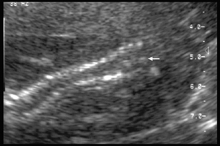
- Bony spur (echogenic focus) through the center of spinal canal.
- Isolated or...
- Associated with other vertebral defects including:
- Spina bifida.
- Kyphoscoliosis.
- Hemivertebra.
- Butterfly vertebra.
- Absent vertebrae.
- Sagittal clefted vertebrae.
- There may be an associated
lipomeningocele or rare teratoma of the cord (3).
- Cutaneous
signs of occult dysraphism include (4):
- Hypertricosis (hairy patch) 56%
-
Capillary hemangioma
26%
-
Dermal sinus 22%
-
Subctaneous lipoma 11%.
- Associations
– High association with a low
lying conus (tethered cord) with thickened filum terminale (40%).
- Hydromyelia of one or both hemicords.
-
Lipoma of the filum terminalis.
-
Intradural lipoma.
- Fibrous bands.
|
|
|
Diastematomyelia |
REFERENCES |
- Silverman FN, Kuhn JP (eds). Caffey’s X-ray pediatric diagnosis: An integrated imaging approach 1993;Mosby St Louis:pg 133-134 and 318-319.
- Pang D. Split cord malformation: Part II. Clinical syndrome. Neurosurgery 1992;31:481-500
- Seeds JW, Powers SK. Early
prenatal diagnosis of familial lipomeningocele. Obstet Gynecol
1988;72:469-471.
- Winter RK, McKnight L, Byrne RA et.al. Diastematomyelia: prenatal ultrasound appearance. Clin Rad 1989;40:291-294
- Sepulveda W, Kyle PM, Hassan J et.al. Prenatal diagnosis of diastematomyelia: case reports and review of the literature. Prenat Diagn 1997;17:161-165.