|
ETIOLOGY /
PATHOGENESIS OF FETAL TRIPLOIDY
|
Most triploid fetuses are lost during the first trimester due to spontaneous abortion. The fetuses that do survive into the second trimester are usually have a wide range of anatomic defects of the head, face heart and extremities, as well as severe asymmetrical growth restriction (1).
A triploid karyotype is present in 90% of cases with partial mole (2-4).
- ± 80% of triploid fertilizations arise from fertilization of a haploid ovum with either a single sperm that reduplicates or two sperm (didandry) (3,5).
- ± 20% are thought to be due to a double maternal contribution (ovum fails to undergo the first or second meiotic division prior to fertilization - digyny) (3,5).
- A supernumerary paternal haploid set is imparted to the ovum resulting in 69 chromosomes with three possible permutations (1):
- XXX
- XXY
- XYY (very few of this karyotype survive to 8 weeks).
Studies suggest that the maternal contribution to the zygote is essential for normal embryonal growth and development, while the paternal contribution is essential for proliferation of extraembryonic tissue (6).
Two phenotypes are described (depending on the parental origin of the extra haploid set) (7,8):
- Type I fetuses ("diandric origin")
- Well grown.
- Proportionate head size (symmetrical growth retardation).
- Partial molar changes in the placenta.
- Placentomegaly.
- Elevated beta hCG
- Increased nuchal translucency (9).
- Type II fetuses ("digynic origin")
- Severe asymmetrical growth retardation.
- No placentomegaly.
- No placental hydatidiform changes.
- <1/3 of second and third trimester triploidy.
- Decreased levels of beta hCG.
- Normal nuchal translucency (9).
|
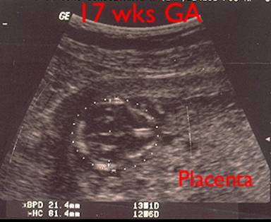
Gestational
age by dates = 17 wks GA by
ultrasound = 13 wks Large cystic placenta |
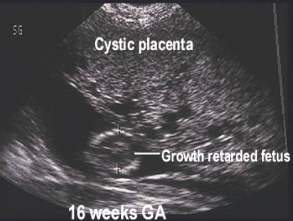
Gestational
age by dates = 16wks GA by
ultrasound = 12 wks Large cystic placenta |
|
|
|
|
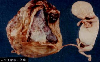
Triploidy
(type II)
|
|
|
|
|

Complications (9) – over 40% of patients are at risk for preeclampsia and a single report of HELLP syndrome has been described).
REFERENCES |
- Dishi N, Surti U, Szulman AE. Morphologic anomalies in triploid liveborn fetuses. Hum Reprod 1983;14:716-723.
- Vassilakos P, Riotton G, Kajii T. Hydatidiform mole: Two entities. A morphologic and cytogenetic study with some clinical considerations. Am J Obstet Gynecol 1977;127:167-170.
- Szulman AE, Surti U. The syndromes of hydatidiform mole. I. Cytogenetic and morphologic correlations. Am J Obstet Gynecol 1978;131:665-671.
- Szulman AE, Surti U. The syndromes of hydatidiform mole. II. Morphologic evolution of the complete and partial mole. Am J Obstet Gynecol 1978;132:22-27.
- Lindor NM, Ney JA, Gaffey TA et.al. A genetic review of complete and partial hydatidiform moles and nonmolar triploidy. Mayo Clin Proc 1992;67:791-799.
- Surani MAH, Barton SC, Norris ML. Nuclear transplantation in the mouse: Hereditable differences between prenatal genomes after activation of the embryonic genome. Cell 1986;45:127-136.
- McFadden DE, Kwong LC, Yam IYL et.al. Prenatal origin of triploidy in human fetuses: Evidence from genomic imprinting. Hum Genet 1993;92:465-469.
- Janiaux E, Brown R, Rodeck C et.al. Prenatal diagnosis of triploidy during the second trimester of pregnancy. Obstet Gynecol 1996;88:983-989.
- Stefos T, Plachouras N, Mari G et.al. A case of partial mole pregnancy and atypical type I triploidy associated wth sever HELLP syndrome at 18 weeks of gestation. Ultrasound Obstet Gynecol 2002;20:403-404.