|
GENERAL FEATURES OF
DOWN SYNDROME |
- Most common chromosomal abnormality (once in every 660 live-births).
- 95% - extra chromosome 21.
3% - due to translocations.
2% - mosaics. - The major abnormalities associated with trisomy 21 are expressed by the genes located at the q22 band of chromosome 21.
- Recurrence risk for trisomy 21 is 1% for women under 30 years of age. Recurrence risk for older patients is the same as the risk associated with their age.
- Table of age specific risk for trisomy 21 in the first and second trimesters and at delivery.
- Maternal serum screening. In trisomy 21, a-fetoprotein levels decrease, urinary E3 levels decrease and bhCG levels increase.
|
Sensitivity and False Positive Rates for Sonographic
Markers in Detection of Second-Trimester Fetuses with Trisomy 21 (1) |
|||
Sonographic Finding
|
Trisomy
21 |
False
positive rate |
Score |
|
Any
major fetal anomaly |
24.5% |
2.8% |
2 |
|
Nuchal Fold 6 mm or greater |
50.9% |
0.6% |
2 |
|
Short
humerus (ratio <0.90) |
41.3% |
3.4% |
1 |
|
Short
femur (ratio <0.91) |
47.2% |
7.9% |
1 |
|
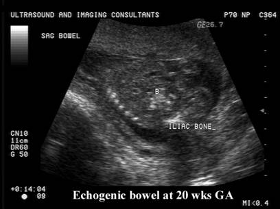
Hyperechogenic bowel |
24.5% |
2.3% |
1 |
|
Echogenic
Intracardiac focus |
30.2% |
4.5% |
1 |
|
Pyelectasis
(AP diameter 4mm or greater) |
22.6% |
0.6% |
1 |
|
Score
of greater than or equal to 1 (risk) |
83% |
17.5% |
|
|
Score
of greater than or equal to 2 (risk) |
75.5% |
5.7% |
|
|
Minor and
major markers of chromosomal aneuploidy |
|
|
|
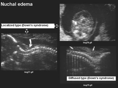
Nuchal edema or fold of above 6mm ·
May result from increased nuchal translucency in the first trimester. ·
Found in 0.5% of fetuses. ·
May be of no pathological significance. ·
May be associated with: o Chromosomal
defects. o Cardiac
anomalies. o Fetal
infection. o Genetic
syndromes. ·
Risk of trisomy
21 in cases of isolated nuchal edema may be 15
times the background risk. |
|
|
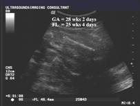
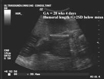
Short femur or humerus
|
|
|
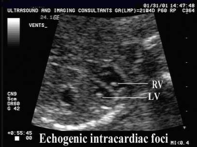
Echogenic intracardiac
foci
|
|
|
Hyperechogenic bowel
|
|
|
Mild pyelectasis (4mm or
greater)
|
|
|
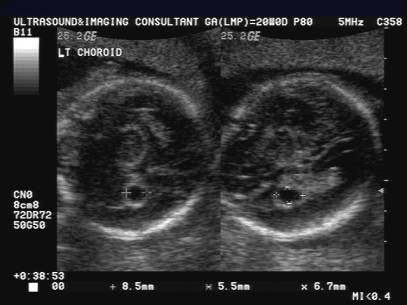
Choroid plexus cysts
|
|
|
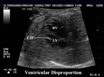
Ventricular Disproportion
|


|
Likelihood
ratios (sensitivity/false-positive rate) of sonographic findings associated
with Down syndrome Nyberg et.al. J Ultrasound Med 2001;20:1053-1063 |
|||
|
Sonographic
marker |
Overall
likelihood ratio (*) |
Likelihood ratio
as an isolated marker (**) |
95% CI |
Nuchal thickening
|
61 |
11 |
5.5 - 22 |
|
Hyperechoic bowel |
33.8 |
6.7 |
2.7 – 16.8 |
|
Short humerus |
15.3 |
5.1 |
1.6 – 16.5 |
|
Short femur |
6.1 |
1.5 |
0.8 – 2.8 |
|
EIF |
6.3 |
1.8 |
1.0 – 3.2 |
|
Pyelectasis |
5.2 |
1.5 |
0.6 – 3.6 |
|
(*) – likelihood ratio when a marker was present either as an isolated finding or in combination with other markers. (**) – likelihood ratio when a marker was present as an isolated finding. EIF – echogenic Intracardiac focus. |
|||
REFERENCES
|
Bromley
B, Lieberman E, Benacerraf BR. The detection of Down
syndrome using a scoring index of sonographic markers and maternal age.
Ultrasound Obstet Gynecol
1997;10:321-324.