|
BREU’S MOLE |
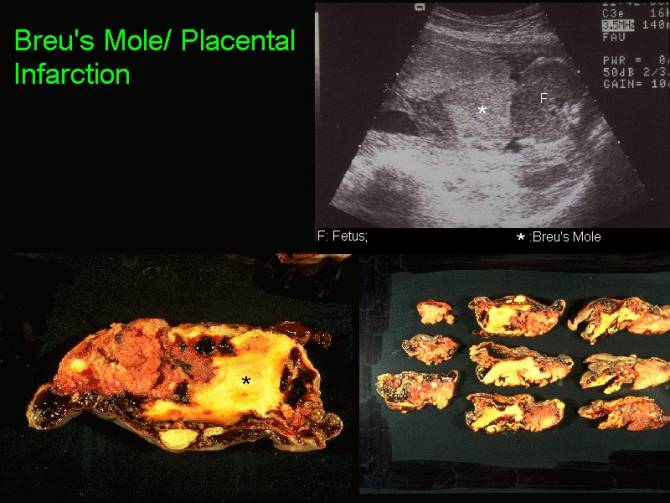
These moles consist of masses of blood thrombus which ultimately forms
fibrin resulting in the term mole (meaning mass) (1).
PATHOLOGY |
These lesions dissect between the layers of the chorionic plate beyond the confines of the intervillous space. The chorionic plate is thus stripped away from the chorionic villous tissue by the thrombohematoma. The clot is formed principally, and probably entirely by maternal blood.
A subchorial thrombohematoma may become intraplacental, which may make the
diagnosis of Breus' mole difficult as it must arise from the chorionic plate to
be classified as a Breus' mole.
ULTRASOUND |
- Thick multilobulated hematoma beneath the chorion.
- Usually confined completely to the subchorial space but may occasionally extend through it.
- Echogenic or hypoechoic depending on the amount of fresh blood present.
- Fetal demise if the placental circulation is affected significantly (1).
|
|
DIFFERENTIAL DIAGNOSIS |
- Hydatidiform mole - little likelihood of confusion.
- Missed abortion - more difficult to differentiate from Breus' mole. Some authors (2) describe Breus mole as a morphologic "variant of missed abortion". The bhCG will probably be higher with Breus' mole as the blood supply of the chorionic tissue is completely severed in missed abortion.
REFERENCES |
- Shanklin DR, Scott JS. Massive subchorial thrombohematoma (Breus' mole). Br J Obstet Gynecol 1975;82:476-487.
- Benirschke K , Driscoll SG. Pathology of the Human Placenta. Spinger, New York 1967;191.