|
SUBAMNIOTIC
HEMORRHAGE - MEMBRANOUS CYST; SUBCHORIONIC CYST; THROMBOTIC CYST |
|
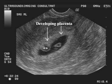
PLACENTAL SURFACE
CYST |
Cystic masses that arise from the fetal surface of the placenta have been referred to by many names )as stated above). Their etiology and clinical importance remains controversial.
Subamniotic hematomas result from the rupture of chorionic vessels (fetal vessels) close to the cord
insertion. These lesions are rarely reported in utero; they are usually
discovered postnatally and thought to result from
excessive traction on the umbilical cord at birth. It has been postulated that
these cysts may form from subchorionic fibrin
deposition (possibly related to X cells in or at their edge).
ULTRASOUND |
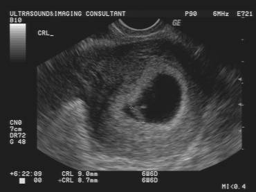
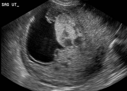
- Usually hypoechoic but may contain echogenic material within the cystic mass. This echogenic area tends to resolve with increasing gestation as the clot resolves.
|
|
|
|
|
|
- Most commonly found near the placental cord insertion.
- Single or multiple.
- It is situated under the amniotic layer covering the fetal (chorionic plate) of the placenta and protrude into the amniotic cavity..
|
|
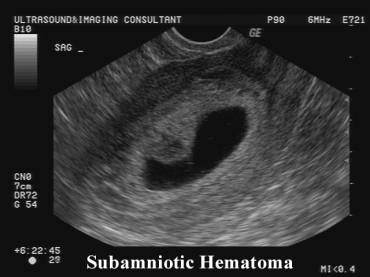
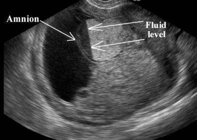
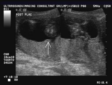
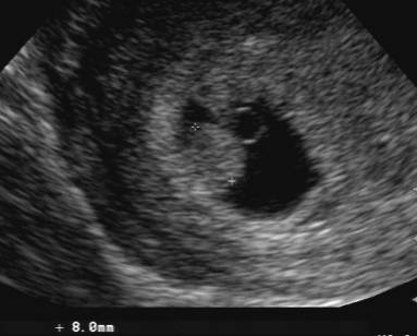
Acute subamniotic hematoma
– hyperechogenic fluid collection (acute blood) situated under the amnion. Note fluid level . Resolevd by 20 weeks. Normal fetal growth until term |
|
|
|
|
|
|
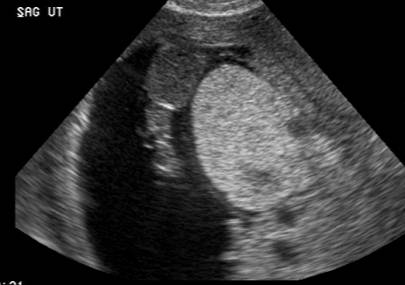
- Single mass.
- Thin covering membrane of amnion over the surface of the mass.
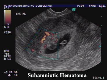
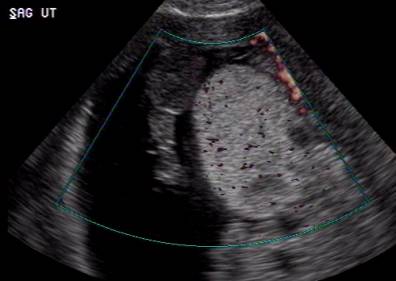
- Avascular on color doppler imaging.
|
|
|
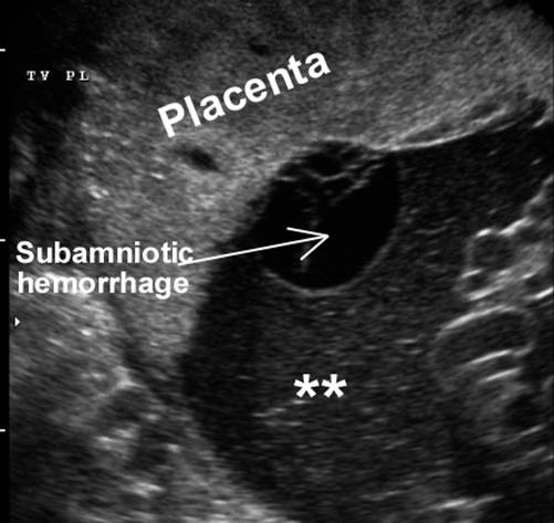
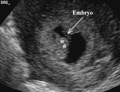
· May rupture into the amniotic cavity resulting in echogenic amniotic fluid (**) due to blood.
|
|
· May present as an echogenic deposit on the fetal surface of the placenta when healed.
|
|
|
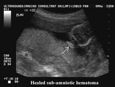
· Usually resolves over time and generally has a good prognosis.
|
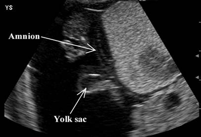
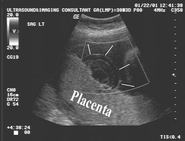
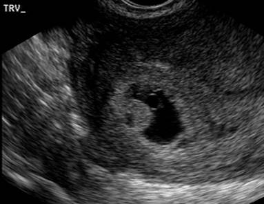
6 weeks of gestation |
|
|
|
|
|
|
|
|
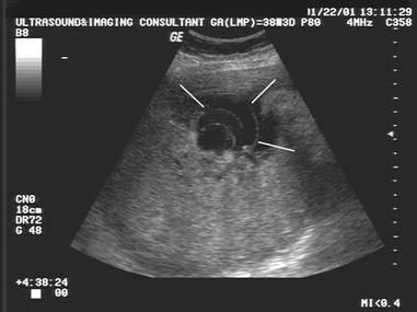
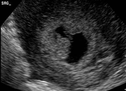
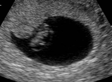
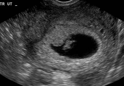
Repeat scan at 7 weeks. Almost
complete resolution of the echogenic hemorrhage. Note the normal development
of the embryo. |
|
|
|
|
COMPLICATIONS
|
IUGR has been reported in approximately 10% of cases (4). Cysts larger than 4.5 cm were associated with IUGR (however 67% of cysts over 4.5 cm were not associated with IUGR). IUGR appeared to be more common when there were more than 3 cysts present.
Maternal floor infarction (MFI) occurred in about 10% of cases (4). This name is a misnomer as the lesions are not infarction but massive fibrinoid deposition in the maternal floor oor basal plate of the placenta. It has been suggested that X cells (trophoblasts outside the villi) may be associated with the formation of these cysts. These X cells occur with increased frequency in MFI and have been found in the walls of these cysts (5).
REFERENCES |
- Kirkinen P, Jouppila P. Intrauterine membranous cyst: a report of antenatal diagnosis and obstetric aspects of two cases. Obstet Gynecol 1986;67:265-305.
- Yiu-Chiu V, Chiu L. Sonographic features of placental complications of pregnancy. Am J Roentgenol 1982;138:850-859.
- Deans A, Jauniaux E. Prenatal diagnosis and outcome of subamniotic hematomas. Ultrasound Obstet Gynecol 1998;11:319-323.
- Brown D, DiSalvo DN, Frates MC et.al. Placental suface cysts detected on sonography. J Ultrasound Med 2002;21:641-646.
- Vernoff KK, Benirschke K, Kephart GM et.al. Maternal floor infarction: relationship to X cells, major basic protein, and adverse perinatal outcome. Am J Obstet Gynecol 1992;167:1355-1362.