|
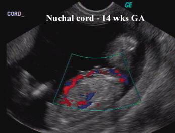
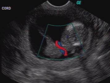
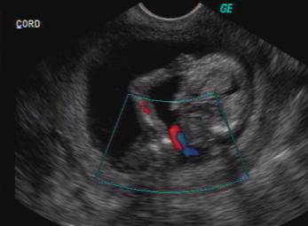
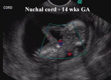
NUCHAL CORD - NECK BODY AND SHOULDER LOOPINGS |
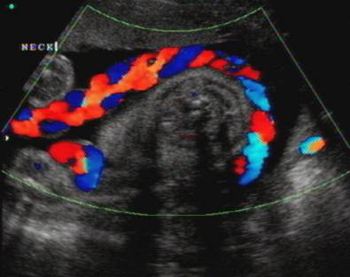
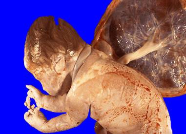
The umbilical cord encircles the fetal neck in about 25% of pregnancies (1).
|
|
|
|
|
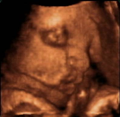
3D view of nuchal cord – single loop |
ULTRASOUND |
- Usually single loops.
|
|
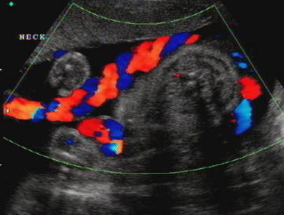
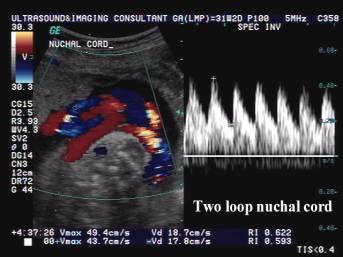
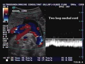
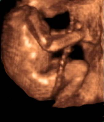
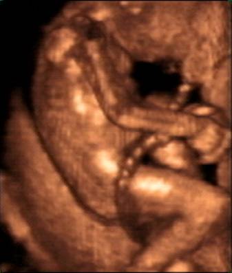
- Multiple loops less frequently:
- 2 loops in 2-3% of cases (1,2).
|
Two loop nuchal cord |
|
|
|
|
- 3 loops in less than 1% of cases (1,2).
- Loops around other parts of the body in 2% of cases.
Cord loop around
upper leg and thigh
|
|
|
|
|
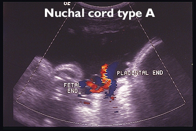
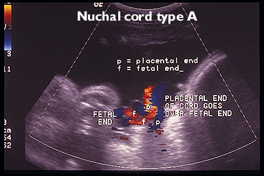
- Nuchal cord type A.
- nuchal loop 360° around the fetal neck where the placental end crosses Over the umbilical end (3).
|
|
|
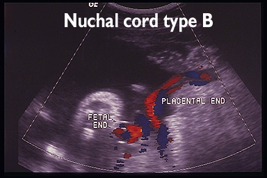
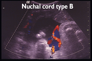
- Nuchal cord type B.
- nuchal loop 360° around the fetal neck where the
placental end crosses Under the umbilical end (3).
Type A is loose and can undo itself whereas type B locks and cannot undo itself (3).
|
|
|
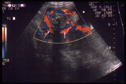
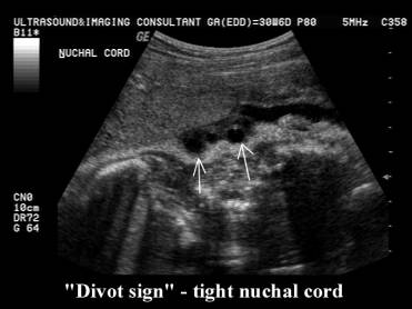
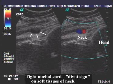
- "Divot sign" (4) - disruption of the smooth contour of the fetal neck compressing skin in that area. Although this sign is described on gray-scale images it may also be demonstrated on color doppler images.
“Divot sign” |
|
|
|
|
|
|
|
|
|
Video clip of
Nuchal Cord – single loop
|
|
|
|
|
|

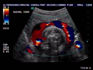
- Color doppler may facilitate ultrasound detection of nuchal cord:
- Jauniaux et.al. (9) assessed sonographic detection of nuchal cord in the third trimester and determined an overall sensitivity of 79% (sensitivity prior to 36 weeks was 67% and after 36 weeks was 93%).
- Funk et.al. (10) found the sensitivity and specificity of diagnosing a nuchal cord during labor was 96% and 97% respectively.
- Single versus multiple loops on color doppler was 72% versus 96% respectively.
- A nuchal cord is more likely to be found at delivery when sonographic examination a few days prior demonstrated a nuchal cord.. Fetal movements can however modify cord position (9).
- Color doppler may demonstrate normal carotid and jugular vessels in the neck. Persistent jugular compression may result in distended facial veins and dural sinus.
- Nuchal cord in the first trimester may affect nuchal translucency measurement.
|
|
|
|
|
|
ASSOCIATIONS |
- Associated with increased cord length, small fetuses, vertex presentation and polyhydramnios (1,2).
- Increased frequency of fetal distress (bradycardia, variable decelerations and depressed 1-minute Apgar scores), when a nuchal cord is present (1,2,4).
- No significant difference in 5-minute Apgar scores nor is there an increase in infant mortality (1,2,5).
- Two or more tight nuchal loops, especially if there is indentation of the skin of the fetal neck by the looped cord, is likely to be associated with an increased fetal mortality (1,2,5).
- Doppler assessment of the cord may reveal high resistance arterial flow or slow venous flow if the loop is hemodynamically significant.
COMPLICATIONS |
- It is uncommon for nuchal cords to result in permanent fetal injury (6,7)
- Entanglement by the fetus and vascular compromise (usually umbilical vein).
- Distended facial veins (8), due to jugular compression.
- Dural sinus distention and ectasia due to protracted jugular pressure (7). This presents as a fluid collection posterior or superior to the cerebellar vermis. Differential diagnosis includes arachnoid cyst, vein of Galen AVM, Dandy-Walker malformation or normal variant). There may be dilatation of the torcular Herophili, vein of Galen, straight sinus and transverse sinus (6).
- Umbilical arterial thrombosis (6).
REFERENCES |
- Miser WF. Outcome of infants born with nuchal cords. J Fam Pract 1992;34:441-444.
- Nyberg DA, Finberg HJ. The
placenta, placental membranes, and umbilical cord. In: Nyberg DA, Mahony
BS et.al. eds. Diagnostic ultrasound of fetal anomalies: text and atlas.
- Giacomello F. Ultrasound determination of nuchal cord in breech presentation. Am J Obstet Gynecol 1988;159:531-532.
- Ranzini AC, Walters CA, Vintzileos AM. Ultrasound diagnosis of nuchal cord: The gray-scale divot sign. Obstet Gynecol 1999;93:854.
- Dudiak CM, Salamon CG, Posniak HV et.al. Sonography of the umbilical cord. Radiographics 1995;15:1035-1050.
- Katz ME, Bass T, White LE. Dural sinus ectasia after prolonged nuchal cord encirclement. J Ultrasound Med 1992;11:289-292.
- Verdel MJC, Exalto N. Tight nuchal coiling of the umbilical cord causing fetal death. J Clin Ultrasound 1994;22:64-66.
- Grimm TW, Cable TA. Nuchal cord: An unusual manifestation. J Am Board Fam Practice 1988;1:218.
- Jauniaux E, Mawissa C, Peellaerts C et.al. Nuchal cord in third trimester pregnancy: a color doppler imaging study. Ultrasound Obstet Gynecol 1992;2:417-419.
- Funk A, Heyl W, Rother r et.al. Subpartal diagnosis of umbilical cord encircirclement using color-coded doppler ultrasonography and correlation with cardiotocographic changes during labor. Geburtshilfe Frauenheilkd 1995;55:623-627.