|
CORD COMPRESSION
(1-9) CORD PRESENTATION AND
PROLAPSE |
Umbilical cord constriction can be due to intrinsic or extrinsic
mechanisms. Constriction may lead to different degrees of flow limitation in
the cord"s vessels, which can be demonstrated by pulsed Doppler flow
studies.
Intrinsic constriction is characterized by localized absence of Wharton"s
jelly, leading to narrowing of the cord, thickening of the vascular walls and
narrowing of the vascular lumens. In this setting, fetal death might occur due
to acute vasospasm, acute oligohydramnios and uterine contraction, or an
obliterating thrombus (10).
Extrinsic constriction can be caused by:
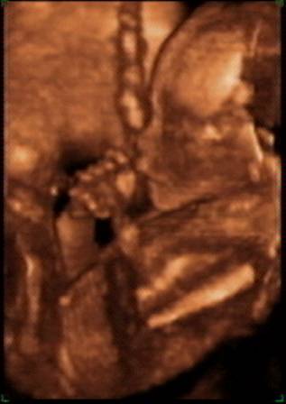
Fetus holding cord at 20 weeks of gestation |
|
|
|
|
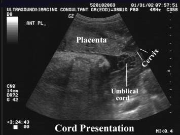
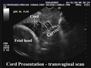
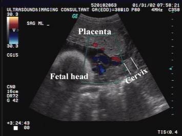
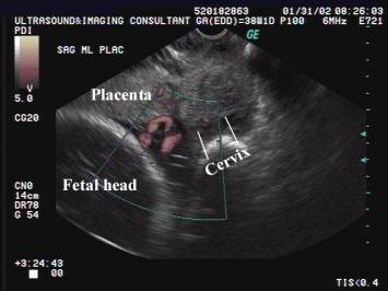
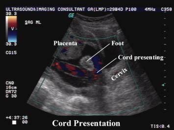
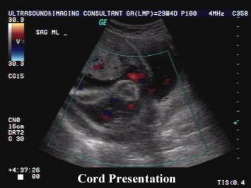
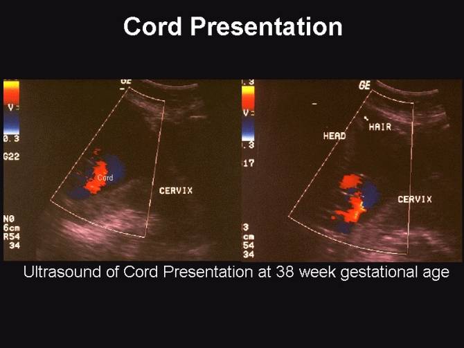
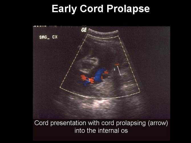
CORD PRESENTATION (1-4) |
Occasionally loops of cord may lie between the lower uterine segment and the presenting part (cord or funic presentation). This is important to recognize as it predisposes to cord prolapse and possible fetal death when the membranes rupture. Funic presentation is more common with malpresentations (especially breech and transverse lie).
CAUSES OF PERSISTENT CORD PRESENTATION (1-4) |
- Transient and usually insignificant prior to 32 weeks. If this is persistent one must look for a cause.
- Marginal cord insertion from the caudal end of a low-lying placenta.
- Uterine fibroids / Uterine adhesions.
- Congenital uterine anomalies that may prevent the fetus from engaging well into the lower uterine segment.
- Cephalopelvic disproportion.
- Polyhydramnios.
- Multiple gestations.
- Increased umbilical cord length.
|
|
|
|
COMPLICATIONS |
- Prolapse of the cord occurs in 0.5% of cases.
- High perinatal mortality rate due to cord compression (1).
REFERENCES |
- Selbing A. Umbilical cord compression diagnosed by means of ultrasound. Acta Obstet Gynecol Scand 1988;67:565-567.
- Hales ED, Westney LS. Sonography of occult cord prolapse. JCU 1984;12:283-285.
- Dudiak CM, Salomon CG, Posniak HV et.al. Sonography of the umbilical cord. Radiographics 1995;15:1035-1050.
- Johnson RL, Anderson JC, Irsik RD et.al. Duplex ultrasound diagnosis of umbilical cord prolapse. J Clin Ultrasound 1987;15:282-284.
- Kanayama MD, Gaffey TA, Ogburn PL Jr. Constriction of the umbilical cord by an amniotic band, with fetal compromise illustrated by reverse diastolic flow in the umbilical artery. A case report. J Reprod Med 1995 Jan;40(1):71-73.
- Boughizane S, Zhioua F, Jedoui A, Kattech R, Gargoubi N, Srasra M, Ben Romdhane K, Meriah S. Swallowing of an amniotic string by a fetus at term. J Gynecol Obstet Biol Reprod (Paris) 1993;22(4):409-410.
- Heifetz SA. Strangulation of the umbilical cord by amniotic bands: report of 6 cases and literature review. Pediatr Pathol 1984;2(3):285-304.
- Reles A, Friedmann W, Vogel M, Dudenhausen JW. Intrauterine fetal death after strangulation of the umbilical cord by amniotic bands. Geburtshilfe Frauenheilkd 1991 Dec;51(12):1006-1008.
- Sherer DM, Anyaegbunam A. Prenatal ultrasonographic morphologic assessment of the umbilical cord: a review. Part I. Obstet Gynecol Surv 1997 Aug;52(8):506-514
- Hallak M, Pryde PG, Qureshi F, Johnson MP, Jacques SM, Evans MI. Constriction of the umbilical cord leading to fetal death. A report of three cases. J Reprod Med 1994 Jul;39(7):561-565.