|
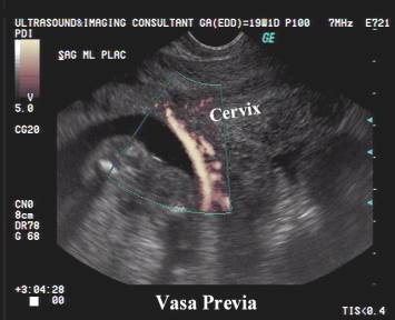
VASA PREVIA |
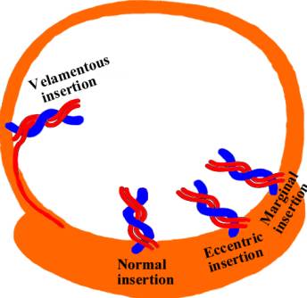
Vasa previa exists when the umbilical vessels of a velamentous insertion traverse the fetal membranes in front of the presenting part. The umbilical arteries and vein are therefore unprotected by placental tissue or umbilical cord. Rupture of membranes may lead to rapid fetal exsanguination as the bleeding is entirely fetal with no significant risk to the mother (except if there is co-existing placenta previa).
There appears to be an increased risk in multiple pregnancies and in pregnancies resulting from in-vitro fertilization.
Lee et.al. (1) describe 18 cases:
· Mean age of diagnosis – 26 weeks (earliest diagnosed at 16 weeks of gestation).
· 8/16 cases had a low-lying placenta on a previous scan and 6 of the patients experienced bleeding complications.
· 3/16 women delivered vaginally as late trimester scans no longer showed vasa previa due to differential growth of the uterus and placenta.
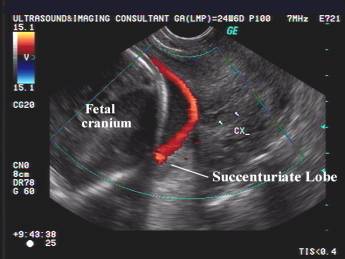
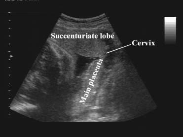
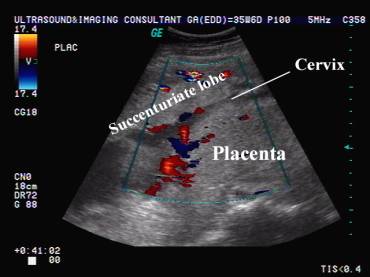
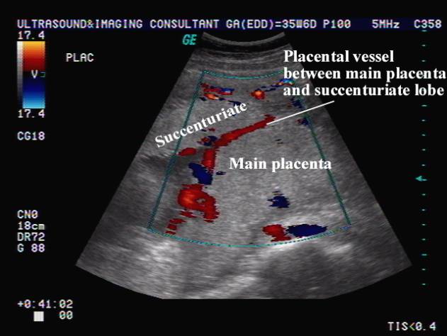
· Risk factors identified for vasa previa included: velamentous insertion (10 patients); bilobed placenta (3 patients); succenturiate placenta lobe (3 patients); multiple gestation (3 sets of twins) and marginal cord insertion (3 patients).
·
The authors recommend at least one additional
scan in the late third trimester to reassess the diagnosis prior to delivery.
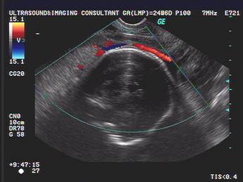
ULTRASOUND |
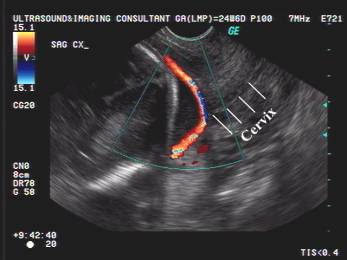
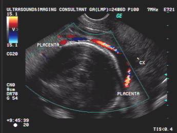
Fetal vessels of the placenta crossing the internal cervical os.
- Vessels connecting separate succenturiate lobe to the main portion of the placenta.
- Cord vessels of velamentous (membranous) cord insertion from a low-lying placenta (prevalence increased in multiple pregnancies).
- Aberrant chorionic vessels in association with marginal cord insertion from a low-lying placenta.
|
|
|
|
|
|
|
|
|
|
|
|
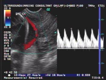
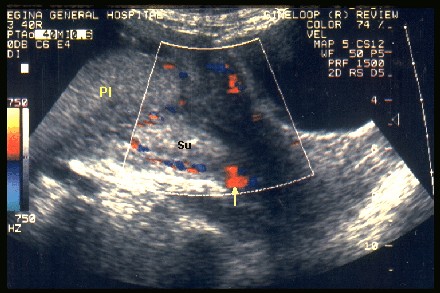
Color and power doppler
defines the relationship of the placental margin, the internal cervical os and the vessels at the margins of the placenta. It
enhances visualization of marginal veins.
|
Complete placenta previa by main placental mass. Succenturiate lobe situated
anteriorly. Aberrant cord vessel between main placental mass and succenturiate lobe. |
|
|
|
|
|
|
|
COMPLICATIONS |
- Perinatal mortality is 50-60% with intact membranes and 70-100% with ruptured membranes.
- Bleeding from torn fetal vessels.
- Cord compression by the presenting part during labor.
- Cord prolapse.
REFERENCES |
- Lee W, Lee VL, Kirk JS et.al. Vasa previa: diagnosis, natural evolution and clinical outcome. Obstet Gynecol 2000;95(4):572-576.
- Hsieh FJ, Chen HF, Ko TM et.al. Antenatal diagnosis of vasa previa by color-flow mapping. J Ultrasound Med 1991;10:397-399.
- Nelson LH, Melone PJ, King M. Diagnosis of vasa previa with transvaginal color Doppler flow imaging. Obstet Gynecol 1990;76:506-509.
- Meyer WJ, Blumenthal L, Cadkin A et.al. Vasa previa: prenatal diagnosis with transvaginal color flow imaging. Am J Obstet Gynecol 1993;169:1627-1629.
- Arts H, van Eyck J. Antenatal diagnosis of vasa previa by transvaginal color doppler sonography. Ultrasound Obstet Gynecol 1993;3:276-278.
- Hata K, Hata T, Fujiwaki R et.al. An accurate diagnosis of vasa previa with transvaginal color Doppler ultrasonography. Am J Obstet Gynecol 1994;171:1265-1267.
- Sauerbrei EE, Davies GL. Diagnosis of vasa previa with endovaginal color doppler and power doppler sonography. Report of two cases. J Ultrasound Med 1998;17:393-398.