|
FETAL FAT
DISTRIBUTION |
Subcutaneous fat, reflected as skinfold thickness has been used to assess neonatal nutritional status, however assessment of fetal fat has been limited (1). The ratio of AC/FL has been suggested to take into account the preferential wasting on the subcutaneous fat tissue over the long bone length among growth-retarded fetuses.
The fetus accumulates most of its body fat during the third trimester (2). Fetal fat stores are second only to the liver weight in reflecting impaired fetal growth (3).
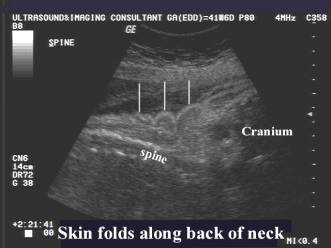
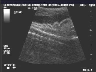
Prior to 24 weeks gestation, very little subcutaneous fat is deposited. After 24 weeks gestation, it is common to see echolucent fat layering the subcutaneous tissues. This is best recognized in the thigh, posterior neck, the malar region and fetal scalp. Skinfolds contain a double layer of skin and subcutaneous tissue.
Total skin fat increases from 4% of body weight at 28 weeks gestation to 14% at 40 weeks gestation (2). At term, 75% of body fat is found in the subcutaneous adipose tissue (4).
Some workers (5) suggest subjective assessment, as measured results are not reproducible. Subcutaneous fat and muscle are indirect indicators of fetal protein and caloric reserves. Newborns with low fat and high muscle reserves have significantly greater birth weight than newborn with high fat and low muscle reserves (6). Evaluation of subcutaneous tissue itself, cannot therefore distinguish normal from impaired growth in utero.
Assessment of fat distribution is most useful in differentiating the normal
small fetus (fat layers are usually present) from the dysmature IUGR fetus (fat
layers are absent, diminished or less widely distributed).
REFERENCES
|
- Winn HN, Holcomb WL. Fetal nonmuscular soft tissue: A prenatal assessment. J Ultrasound Med 1993;4:197-199.
- Widdowson EM, Southgate DAT, Hey EN. Body composition of the fetus and infant. In: Vesser HKA (ed). Nutrition and metabolism of the fetus and infant. The Hague. Martinus Nijhoff Publishers 1979:169-177.
- Brans YW, Cassady G. Intra-uterine growth and maturation in relation to fetal deprivation. In: Gruenwald P (ed). The placenta. University Park Press, Baltimore 1975:307-334.
- Dauncey MJ, Gandy G, Gairdner D. Assessment of total body fat in infancy from skinfold thickness measurements Arch Dis CHILD 1977;52:223-227.
- Hill LM, Guzick D, Boyles D et.al. Subcutaneous tissue thickness cannot be used to distinguish abnormalities of fetal growth. Obstet Gynecol 1992;80:268-271.
- Frisancho AR, Klayman JE, Maltos J. Newborn body composition and its relationship to linear growth. Am J Clin Nutr 1977;30:704-711.