|
|
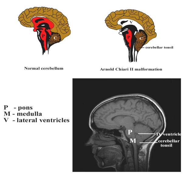
The Arnold-Chiari malformation is a defect in which the brainstem is drawn down into the foramen magnum due to tethering and traction of the spinal cord (usually due to an open spinal defect). The brain herniation results in external compression of the IV ventricle, which in turn disrupts normal CSF circulation resulting in obstructive hydrocephalus.
- Present in almost every case of thoracolumbar, lumbar and lumbosacral myelomeningocele.
- Inferior displacement of the medulla and fourth ventricle into the upper cervical canal.
- Elongation and thinning of the upper medulla and lower pons and persistence of the embryonic flexures of these structures.
- Hydrocephalus.
ETIOLOGY
|
- The Chiari II malformation is due to in utero leakage of CSF through the open neural tube defect.
- Leaking of CSF out of the central nervous system doe not allow the fourth ventricle to expand (due to lack of normal hydrostatic pressure).
- The fourth ventricle is too small and the posterior fossa fails to develop normally.
- Because the posterior fossa is too small, the growing brainstem and cerebellum herniated outside the confines of the posterior fossa.
- The pons and fourth ventricle are displaced inferiorly, forming a cervicomedullary kink within the cervical canal.
- The cerebellar tonsils herniated through the foramen magnum and for a “peg” behind the cervical cord.
- Anterior extension of the cerebellum causing petrous scalloping, upward cerebellar protrusion through the tentorium (the “towering” cerebellum), tectal beaking of the midbrain, dysplasia of the corpus callosum, hypoplasia of the falx cerebri, and stenogyria (abnormal gyral folding) (2).
|
|
|
|
|
VentriculomegalyMinimal lemon
sign Banana
cerebellum Obliterated cisterna magna |
|
|
|
|
|
|
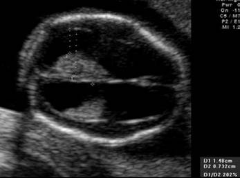
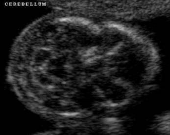
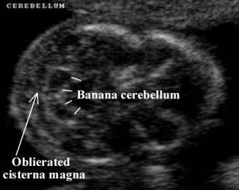
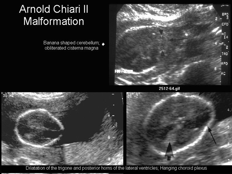
- Inferior displacement of the inferior cerebellum through the foramen magnum (Banana sign).
- Lemon
sign
- Bony defects of the foramen magnum, occiput and upper cervical vertebrae.
- Hydrocephalus from the hindbrain malformation that blocks the flow of CSF through the fourth ventricle or the posterior fossa, or from aqueductal stenosis which may be present in 40-75% of cases.
- Clivus-supraocciput angle has been found to be a useful parameter in differentiating the various causes of ventriculomegaly, particularly Chiari II malformations (1). The angle is measured in a mid sagittal plane of the fetal skull between the clivus and supraocciput. It is constant during gestation and measures 79.3 degrees +/- 6 degrees. Chairi II malformations tend to have values below the 10th percentile.
Clivus-supraocciput angle in fetal ventriculomegaly |
||
Cause of ventriculomegaly |
GA (wks) |
Mean angle
(degrees) |
|
Chiari II malformation (13 cases) |
16-34 wks |
65.1 |
|
Agenesis of the corpus callosum (12 cases) |
25-34 wks |
80.6 |
|
Aqueductal stenosis |
17-26 wks |
81.5 |
|
Borderline ventriculomegaly |
25-34 wks |
80.1 |
|
Dandy-Walker malformation |
20-23 wks |
80.3 |
|
Porencephaly |
25-33 wks |
84.5 |
|
Schizencephaly |
33 wks |
85 |
ASSOCIATED ANOMALIES |
- Spinal anomalies.
- Lumbar myelomeningocele (>95%).
- Syringohydromyelia.
- Supratentorial anomalies.
- Dysgenesis of the corpus callosum (80-95%).
- Obstructive hydrocephalus (50-98%) following closure of meningomyelocele.
- Absent septum pellucidum (40%).
- Polymicrogyria.
REFERENCES |
- Addario VD, Pinto V, Di Naro E et.al. The
posterior fossa: a useful landmark in the evaluation of fetal ventriculomegaly. 10th World Congress on
Ultrasound in Obstetrics and Gynecology 2000:
- McLone DG, Naidich TP. Developmental morphology of the subarachnoid space, brain vasculature, and contiguous structures, and the cause of the Chiari II malformation. Am J Neuroradiol 1992;13:463-482.