|
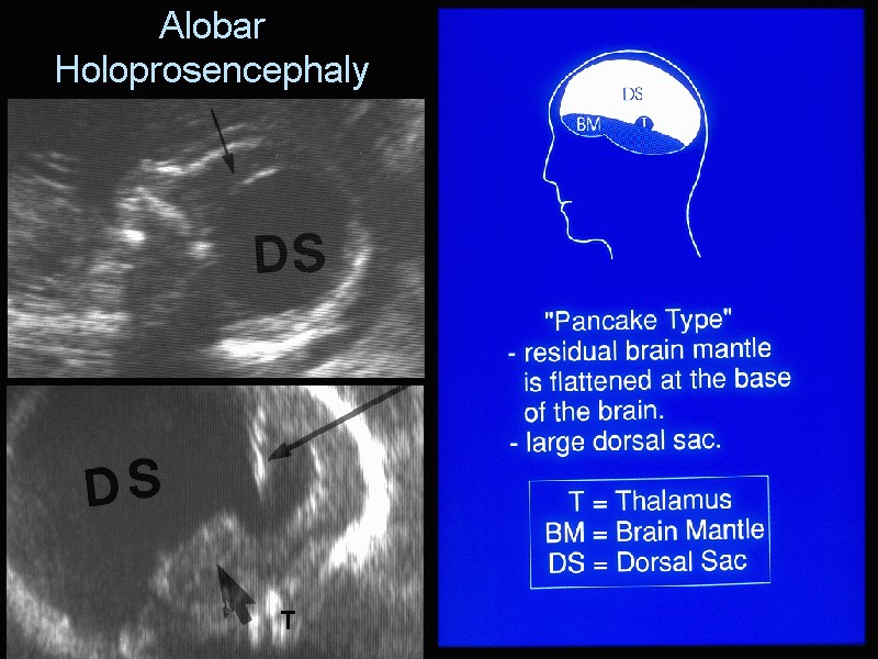
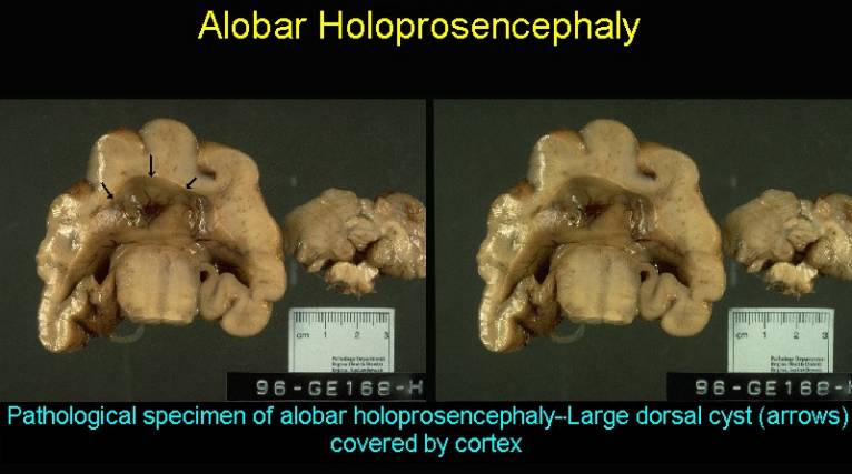
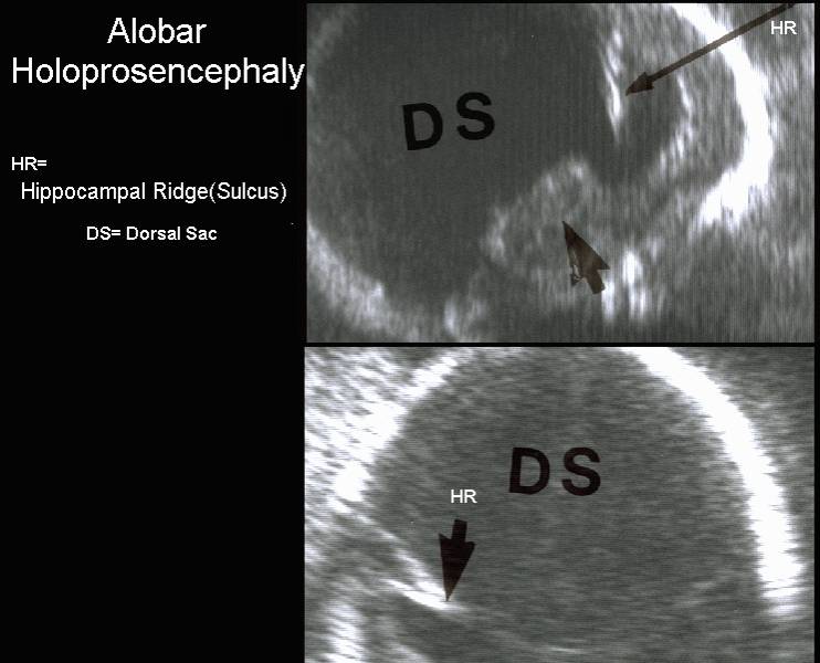
ALOBAR
HOLOPROSENCEPHALY |
This is the most severe form in which the prosencephalon
fails to divide.
ULTRASOUND |
- Interhemispheric fissure absent.
- Falx cerebri absent.
- Single primitive ventricle (holoventricle) with a large dorsal cyst.
- "Horseshoe" or "boomerang" configuration of the brain (peripheral rim of cerebral cortex displaced rostrally in a coronal plane).
- Pancake configuration - cortex covers monoventricle to edge of dorsal cyst.
- Cup configuration = more cortex present posteriorly.
- Ball configuration = complete covering of monoventricle with dorsal cyst.
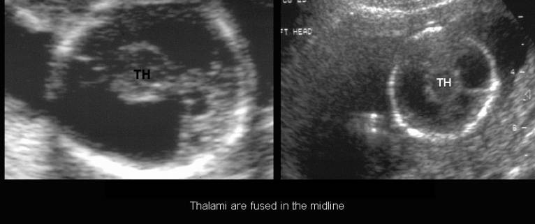
- Thalami are fused in the midline (thalami and basal ganglia protrude into the monoventricle).
- Midbrain, brainstem and cerebellum are structurally normal.
- Absent third ventricle, neurohypophysis, olfactory bulbs and tracts.
- Absent septum pellucidum, falx cerebri and corpus callosum.
|
|
|
|
|
|
|
|
ORBITAL AND FACIAL ANOMALIES |
(5 categories described) (1).
A normal face is present in 17% of cases.
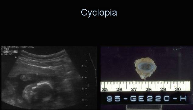
- Cyclopia.
- 1 eye or partially divided eyes in a single orbit.
- Arhinia (absent nose with a proboscis that implants above the orbit).
|
|
|
|
|
|
|
|
|
|

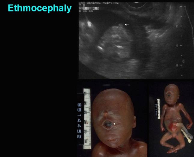
- Ethmocephaly.
- Marked hypotelorism, arhinia, absent nose with a proboscis above the orbit or between narrowly placed orbits.
|
|
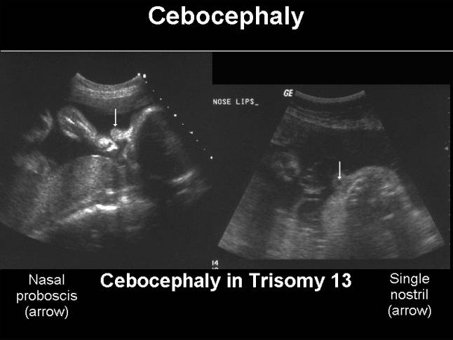
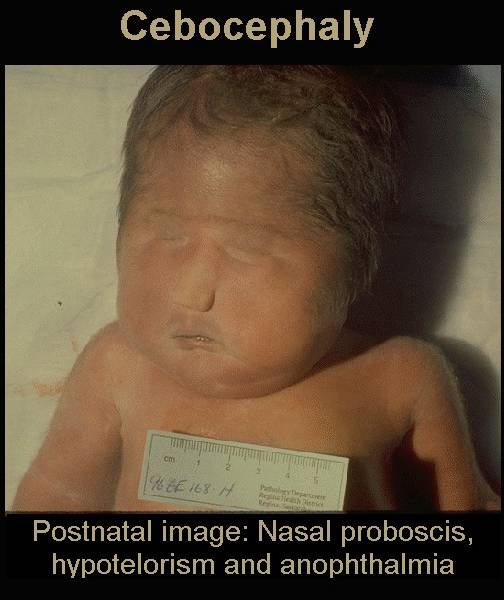
- Cebocephaly.
- Marked hypotelorism, proboscis like nose (normally placed but having a single nostril).
|
|
|
|
- Face with median cleft
lip.
- Hypotelorism with a flattened or absent nose.
- Face with median philtrum-premaxilla a large and flat nose.
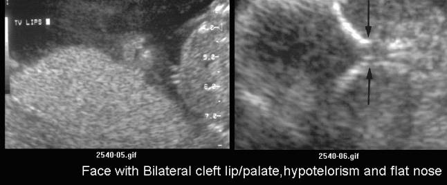
- Bilateral cleft lip/palate, hypotelorism and flat nose.
|
|
|
1+2 |
- Alobar holoprosencephaly |
Other less common facial anomalies include micrognathia,
trigonocephaly, microphthalmia
and microcephaly.
EXTRAFACIAL ANOMALIES ASSOCIATED WITH HOLOPROSENCEPHALY |
Extrafacial anomalies have been reported in approximately 52% of cases (2)
- Myelomeningocele.
- Renal dysplasia.
- Omphalocele.
- Esophageal atresia.
- Cardiac defects.
REFERENCES |
- DeMyer W, Zeman W. Alobar holoprosencephaly (arhinencephaly) with median cleft lip and palate: Clinical electroencephalographic and nosologic considerations. Confin Neurol 1963;23:1.
- McGahan
JP, Nyberg DA,