|
LOBAR
HOLOROSENCEPHALY |
The fetal brain is almost completely divided into two distinct hemispheres
except for a variable degree of fusion at the level of the cingulate gyrus and
frontal horns of the lateral ventricle. The prognosis is uncertain. May have a
normal life span but mental retardation and neurological sequelae are common.
ULTRASOUND (1-9) |
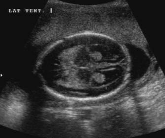
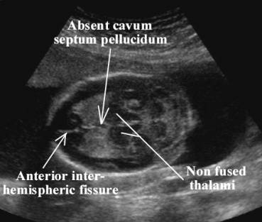
- Absent cavum septum pellucidum (100%).
|
|
|

- Corpus callosum and olfactory bulbs and tract (absent/normal/hypoplastic).
|
|
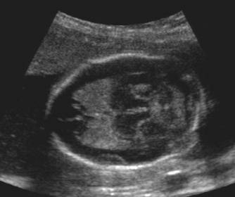
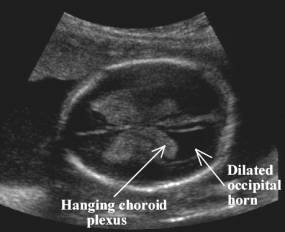
Colpocephaly with dilatation of the occipital horns due to callosal
agenesis |
|
|
|
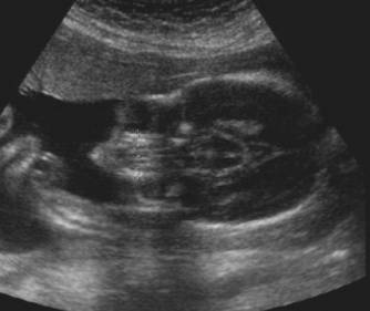
- Squaring of the roof of the frontal horns of the lateral ventricle.
- Incomplete segregation of frontal horns and 3rd ventricle (fused frontal horns communicate centrally with a slightly enlarged 3rd ventricle).
- Intraventricular fused fornices in the cavity of the 3rd ventricle. A "thick fascicle", representing the fornices abnormally fused in the midline, is present within the fused lateral and 3rd ventricle. It runs in the midline between the anterior and posterior commisure.
|
|
|
|
|
|
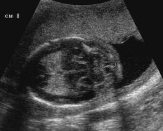

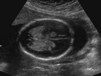
- Incomplete / complete interhemispheric fissure.
|
|
|
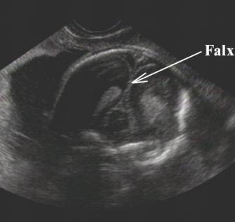
- Falx cerebri - hypoplastic or absent.
- Thalami rounded (bulb shaped).
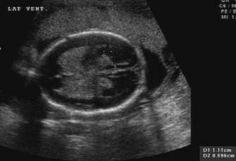
- Borderline ventriculomegaly (10-15mm) in the majority of fetuses early on. Overt hydrocephalus usually occurs later in pregnancy and is thought to be due to dysplasia of the aqueduct of Sylvius.
- Orbits and interorbital distance is normal.
|
|
·
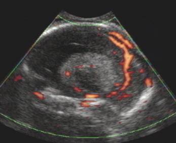
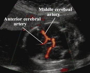
Vascular anomalies associated with lobar
holoprosencephaly (6-9).
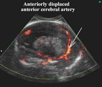
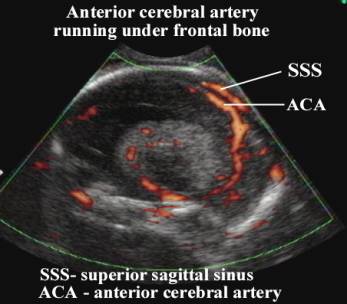
The abnormal trajectory of the anterior cerebral artery in cases of holoprosencephaly has been well described. Lobar holoprosencephaly always has fusion of the two frontal gyri (even if it is only partial). The anterior cerebral artery is pushed externally alongside the frontal bone by the abnormal bridge of cortical tissue between the two frontal gyri. This has been described as the “snake under the skull: appearance on sagittal views of the brain when asses with color or power Doppler.
|
|
|
|
|
Anterior displacement of the anterior cerebral artery on
the sagittal images. |
|
|
Displacement of the anterior cerebral artery cannot be
appreciated on the axial images. |
ASSOCIATED MALFORMATIONS |
- Dandy Walker Malformation.
- Pachygyria or lissencephaly.
|
|
|
DIFFERENTIAL DIAGNOSIS |
REFERENCES
|
- Wong HS, Lam YH, Tang
MH, Cheung LW, Ng LK, Yan KW. First-trimester ultrasound diagnosis of
holoprosencephaly: three case reports. Ultrasound Obstet Gynecol 1999; 13:
356-359
- Tongsong T, Wanapirak C, Sirichotiyakul S, Siriangkul S. First trimester sonographic diagnosis of holoprosencephaly. J Med Assoc Thai 1998; 81: 208-213
- Peebles DM. Holoprosencephly. Prenat Diagn 1998; 18: 477-480
- Turner CD, Silva S, Jeanty P. Prenatal diagnosis of alobar holoprosencephaly at 10 weeks of gestation. Ultrasound Obstet Gynecol 1999; 13: 360-362
- Cohen MM Jr. Perspectives on holoprosencephaly. Teratology 1989; 40: 211-235
- Van Overbeeke JJ, Hillen B, Vermeij-Keers C. The arterial pattern at the base of arhinencephalic and holoprosencephalic brains. J Anat 1994; 185: 51-63
- Arnold WH, Sperber GH, Machin GA. Anatomy of the circle of Willis in three cases of human fetal synophthalmic holoprosencephaly. Anat Anz 1996; 178: 553-558
- Osaka K, Sato N, Yamasaki S, Fujita K, Matsumoto S. Dysgenesis of the deep venous system as a diagnostic criterion for holoprosencephaly. Neuroradiology 1977; 13: 231-238
- Maki K, Kumagai K. Angiographic
features of alobar holoprosencephaly. Neuroradiology 1974; 6: 270-276