|
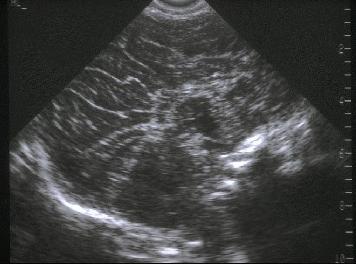
ULTRASOUND OF CALLOSAL AGENESIS -
Complete agenesis -
Partial agenesis |
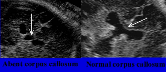
The corpus callosum is a thin band of white matter, which is difficult to
demonstrate sonographically. It is only well visualized on mid-sagittal or
mid-coronal views of the brain and requires optimal angles of insonation to
demonstrate.
COMPLETE AGENESIS OF THE CORPUS CALLOSUM |
|
|
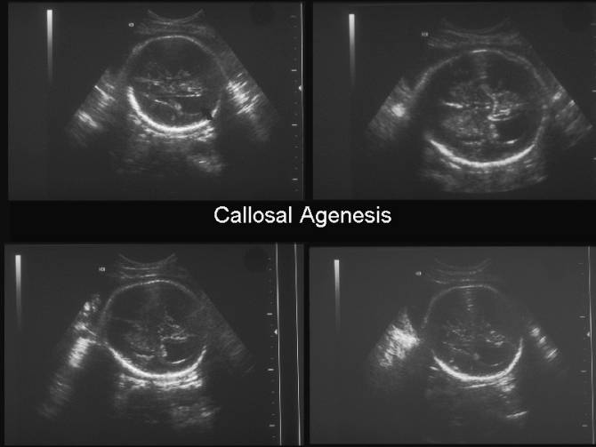
- Absent septum pellucidum.
- Absent corpus callosum and cavum septi pellucidi.
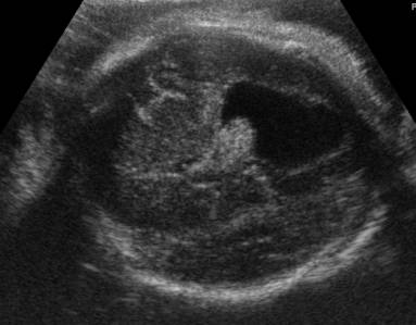
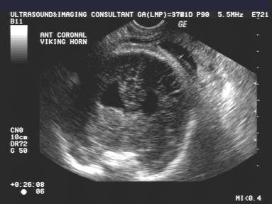
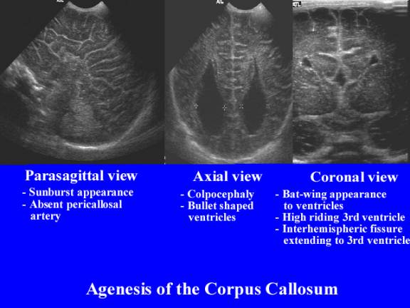
- Teardrop or crescentic shape to the lateral ventricles due to the longitudinal bundles of Probst. The ventriculomegaly is stable and is not associated with intracranial hypertension.
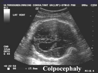
- Colpocephaly (dilatation of the trigones, occipital and temporal horns in the absence of the splenium).
- "Bat-wing" appearance of the lateral ventricles (wide separation of the lateral ventricles with straight parallel parasagittal orientation, with absent body).
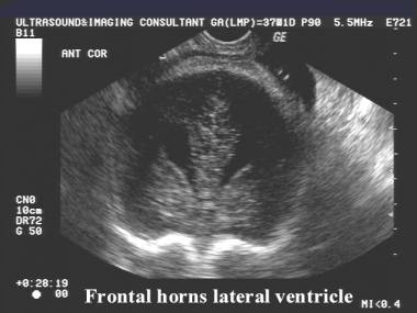
- Laterally convex frontal horns (absent genu of the corpus callosum).
- Frontal horns - normal size but more separated from the midline.
|
|
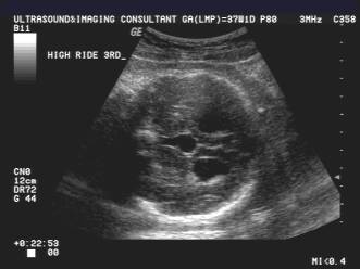
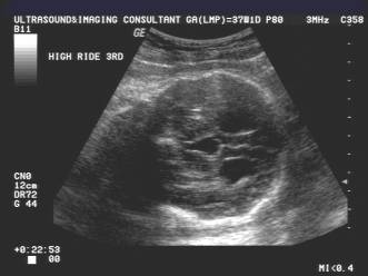
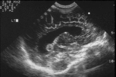
Interhemispheric cyst,high-riding 3rd ventricle |
|
|
Sunburst appearance of gyri |
|
|
Interhemispheric cyst,high-riding 3rd ventricle |
|
|
Colpocephaly |
|
|
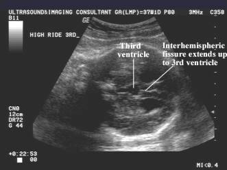
Interhemispheric fissure adjoins the 3rd ventricle |
|
|
Colpocephaly (dilatation of the occipital horn of the
lateral ventricle). |
|
|
|
|
|
|
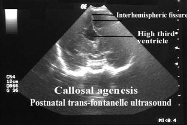

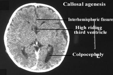
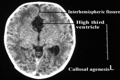
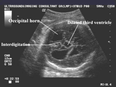
- Third ventricle - "high-riding" (upward displacement of a widened third ventricle often up to the level of the body of the lateral ventricle).
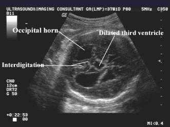
- Anterior interhemispheric fissure adjoins the elevated third ventricle ± communication (Pathognomonic).
- "Interhemispheric cyst" (interhemispheric CSF collection as an upward extension of the third ventricle).
- Enlarged foramen of Munro.
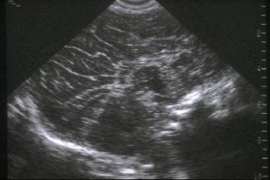
- Sunburst pattern of the gyri (dysgenesis of the cingulate gyrus with characteristic radial orientation of the sulci from the roof of the third ventricle, as seen on a mid sagittal image).
|
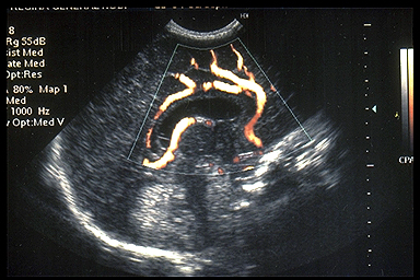
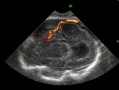
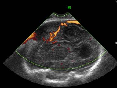
Callosal Agenesis – absent cingulated gyrus and pericallosal artery |
Normal Corpus Callosum – normal pericallosal artery |
|
|
|
|
|
|

- Failure of normal convergence of the calcarine and parieto-occipital sulci.
- Color Doppler - absent pericallosal artery (normally runs along the superior surface of the corpus callosum in a semicircular loop).
|
Normal cingulate gyral pattern. |
Dysgenesis of the cingulate gyrus with characteristic radial
orientation of the sulci |
|
|
|
|
Normal corpus callosum, pericallosal (PC) and
callosomarginal (CM)arteries. |
Agenesis of the corpus callosum with a
"sunburst" gyral pattern and non visualization of the pericallosal
or callosomarginal arteries |
|
|
|
- Posterior fossa - normal cerebellum and cisterna magna.
|
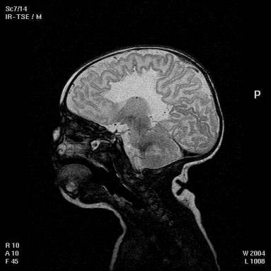
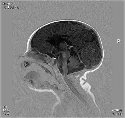
Postnatal MRI scan |
|
|
|
|
Etiology:
o
Lipoma
or interhemispheric cysts (preventing the progression of callosal axons as they
are median structures) (7).
o
Late
remodelling abnormality of callosal connections causing thinning of the corpus
callosum may occur in utero or even at birth after severe asphyxia.
o
Prenatal
ultrasound examination can usually diagnose CACC from 22 weeks onwards
(8).
o
Absence
of the cavum septi pellucidi, upward displacement and enlargement of the third
ventricle
o
Enlarged
occipital horns (colpocephaly) of the lateral ventricles,
o
separated
and enlarged anterior horns,
o
enlarged
interhemispheric spaces are the key indirect signs of CACC.
o
Color
Doppler can also show the loss of the semicircular loop of the pericallosal
artery (8)
PARTIAL AGENESIS (DYSGENESIS) OF THE CORPUS CALLOSUM |
The caudal portion of the corpus callosum (splenium and body) are missing to various degrees.
Partial agenesis affects
only the posterior part of the corpus callosum.
o Indirect signs are lacking and prenatal diagnosis is therefore more difficult (9).
o Hypoplasia occurs as a result of late destruction of the corpus callosum (10) owing to a metabolic, infectious or ischemic origin.
|
Amino acid metabolism |
non-ketotic hyperglycinemia |
|
maternal phenylketonuria |
|
|
methyl malonic acidemia |
|
|
Mitochondrial |
pyruvate dehydrogenase
deficiency |
|
pyruvate decarboxylase deficiency |
|
|
fumarase deficiency |
|
|
Peroxisomal |
Zellweger syndrome |
|
Refsum syndrome |
|
|
adrenoleukodystrophy |
|
|
Other metabolic |
glutaric acidemia |
|
congenital disorder of
glycosylation |
|
|
3-hydroxyisobutyric aciduria |
|
|
Ante or postnatal ischemic |
necrotizing enterocolitis |
|
severe birth asphyxia |
|
|
antenatal cerebral vascular
infarction |
|
|
fetal toxoplasmosis or rubella |
|
|
Chromosomal |
trisomy 8, 13, 18, 21 |
|
Other syndromes |
Menkes syndrome |
|
Smith-Lemli-Opitz syndrome |
|
|
Shapiro syndrome |
|
|
fetal alcohol syndrome |
|
|
acrocallosal syndrome |
|
|
septo-optic dysplasia |
|
|
ectodermal dysplasia |
|
|
Other cerebral |
defect of gyration pattern,
microgyria |
|
microcephaly |
|
|
anomaly of cerebral white matter |
|
|
cerebellar dysplasia |
|
|
interhemispheric cyst |
|
|
Paupe A, Bidat L, Sonigo P et.al. Prenatal diagnosis of
hypoplasia of the corpus callosum in association with non-ketotic
hyperglycemia.
Ultrasound Obstet Gynecol 2002;20:616-619 |
|
REFERENCES |
- Pilu G, Sandri F, Perola A et.al. Sonography of fetal agenesis of the corpus callosum: A survey of 35 cases. Ultrasound Obstet Gynecol 1993;3:318.
- Gebarski SS, Gebarski KS, Bowerman RA et.al. Agenesis of the corpus callosum. Sonographic features. Radiology 1984;151:443.
- Babcock DS. The normal, absent and abnormal corpus callosum. Radiology 1984;151:449.
- Vergani P, Ghidini A, Mariani S et.al. Antenatal sonographic findings of agenesis of the corpus callosum. Am J Perinatol 1988;8:105.
- Lockwood CJ, Ghidini A, Aggarwal R et.al. Antenatal diagnosis of partial agenesis of the corpus callosum. A benign cause of ventriculomegaly. Am J Obstet Gynecol 1988;159:184.
- Hilpert PL, Kurtz AB. Prenatal diagnosis of agenesis of the
corpus callosum using endovaginal ultrasound. J Ultrasound Med 1990;9:363.
- Wrainwright H, Bowen R, Radcliffe M. Lipoma of corpus callosum associated with dysraphic lesions and trisomy 13. Am J Med Genet 1995; 57: 10-3
- D'Ercole C, Girard N,
Cravello L, Boubli L, Potier A, Raybaud C, Blanc B. Prenatal diagnosis of
fetal corpus callosum agenesis by ultrasonography and magnetic resonance
imaging. Prenat Diagn
1998; 18: 247-53
- Tepper R, Zalel Y, Gaon E, Fejgin M, Beyth Y. Antenatal
ultrasonographic findings differentiating complete from partial agenesis
of the corpus callosum. Am J
Obstet Gynecol 1996; 174: 877-8
- Schaeffer GB, Bodensteiner JB, Thompson JN, Wilson DA. Clinical and morphometric analysis of the hypoplastic corpus callosum. Arch Neurol 1991; 48: 933-6
- Malinger G, Zakut H. The corpus callosum: normal fetal development as shown by transvaginal sonography. AJR Am J Roentgenol 1993; 161: 1041-3