|
ULTRASOUND IN
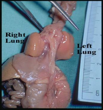
PULMONARY HYPOPLASIA |
The fetal thorax grows at a regular rate from 16 to 40 weeks, resulting in a linear correlation between gestational age and thoracic size.
- Normal thoracic
circumference
- Normal thoracic length
- Ratios between abdominal circumference, head circumference, BPD and femur length remain constant with a high correlation coefficient in normal pregnancy (1,2).
Thoracic circumference / Abdominal circumference is normally >0.8
after 20 weeks. There is a high positive predictive value for pulmonary
hypoplasia using chest circumference measurements (3).
ULTRASOUND |
- Ultrasound (coarsening of echotexture / progressive increase in sound transmission) is a poor predictor of lung maturity (4,5).
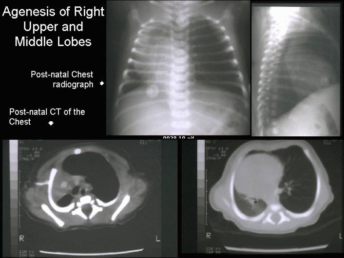
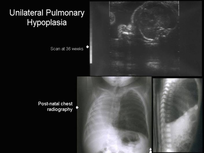
- Unilateral and
involve one or more lung segments/
- Unilateral
involvement of the entire lung.
- Bilateral (Potters Sequence)
|
|
|
|
|
|
|
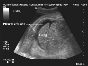
Pleural effusion – no pulmonary hypoplasia |
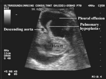
Pleural effusion – pulmonary hypoplasia |
|
|
|
|
Bilateral pulmonary hypoplasia |
|
- Ratio between biventricular outer dimension and the thoracic diameter.
- Constant throughout the second and third trimester.
- Biventricular outer dimension = transverse diameter of the heart at the level of the atrioventricular valves measured at end diastole, increases linearly with advancing age.
- An increased cardiothoracic ratio may result from cardiomegaly without pulmonary hypoplasia or small lungs with a normal heart. One therefore needs to determine which organ system is abnormal in size (6).
- Deviation of cardiac position or axis (7).
- May result from either an intrathoracic mass or an abnormality of the thoracic cage. Both conditions have a high incidence of pulmonary hypoplasia.
- Fetal breathing.
- The implications of fetal breathing activity remains controversial with respect to the prenatal diagnosis of pulmonary hypoplasia and the prediction of fetal outcome.
REFERENCES |
- Fong K, Ohlsson A, Zalev A. Fetal thoracic circumference: A prospective cross sectional study with real time ultrasound. Am J Obstet Gynecol 1988;158:1154-1160.
- Nimrod C, Nicholson S, Davies D et.al. Pulmonary hypoplasia testing in clinical obstetrics. Am J Obstet Gynecol 1988;158:277-280.
- D'Alton M, Mercer B, Riddick E et.al. Serial thoracic versus abdominal circumference ratios for the prediction of pulmonary hypoplasia in premature rupture of membranes remote from term. Am J Obstet Gynecol 1992;166:658-663.
- Fried AM, Loh RK, Umer MA et.al. Echogenicity of fetal lung: Relation to fetal age and maturity. AJR 1985;145:591-594.
- Cayea PD, Grant DC, Coublet PM et.al. Prediction of fetal lung maturity: Inaccuracy of study using conventional ultrasound instruments. Radiology 1985;155:473-475.
- DeVore GR, Horenstein J, Platt LD. Fetal echocardiography: Assessment of cardiothoracic disproportion - A new technique for the diagnosis of thoracic hypoplasia. Am J Obstet Gynecol 1986;155:1066-1074.
- Comstock CH. Normal fetal heart axis and position. Obstet Gynecol 1987;70:255-259.