|
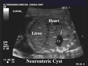
NEURENTERIC CYST |
Neurenteric cysts are posterior enteric remnants that result from incomplete separation of the notocord from the foregut in the third week of embryogenesis. The cyst wall contains both gastrointestinal and neural elements and connects to the meninges.
This is thought to result from a persistent communication or adhesion (1) between the ectoderm of the spinal cord and the endoderm of the foregut before neural tube closure. The adhesion that prevents separation may be caused by an inadequate nutrient supply of the neural folds.
There is a high association of concomitant vertebral anomalies, which determine the prognosis. These anomalies usually occur more cephalad than the cystic chest mass because of differential rates of longitudinal growth of the spine and thorax (2).
Although the cyst commonly attaches to a portion of the gastrointestinal
tract, it rarely communicates with the gut lumen within the thorax.
ULTRASOUND |
- Sites of occurrence:
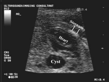
- Posterior mediastinum of the chest (usually on the right side superior to the carina) – 90%.
- In the spinal canal (usually the lower cervical and upper thoracic regions.
- Cystic mass in the chest (1). Cyst size does not appear to correlate with prognosis.
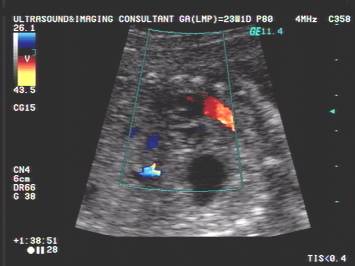
- May displace the lungs, heart airways and great vessels.
- Pulmonary hypoplasia may occur if the mass or displacement is great.
- Fetal hydrops may occur (may be due to mediastinal shift and central venous compression and obstruction).
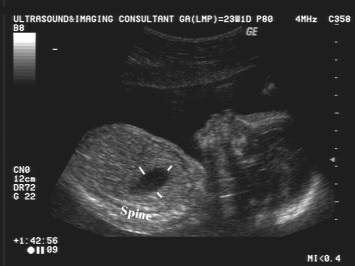
- Spinal anomalies (located at the same level or cranial to the cyst) – 50%:
- Scoliosis.
- Spina bifida.
- Hemivertebrae.
- Butterfly vertebrae.
|
|
|
|
|
|
DIFFERENTIAL DIAGNOSIS OF A CYSTIC CHEST MASS |
- Macrocystic cystic adenomatoid malformation.
- Congenital diaphragmatic hernia.
- Bronchogenic cyst.
- Neurenteric cyst.
The presence of associated vertebral anomalies suggests the latter
diagnosis.
REFERENCES |
- Macaulay K, Winter III TC, Shields LE. Neurenteric cyst shown by prenatal sonography. AJR 1997;169:563-565.
- Haddon M, Bowen A.
Bronchopulmonary and neurenteric forms of foregut anomalies: imaging for
diagnosis and management. Radiol Clin North Am 1991;29:241-254.
- Uludag S, Madazli R, Erdogan E et.al. A case of prenatally diagnosed fetal neurenteric cyst. Ultrasound Obstet Gynecol 2001;18:277-279.