|
NORMAL AND ABNORMAL
CARDIAC POSITION - SITUS ANOMALIES |
NORMAL CARDIAC POSITION |
Abnormal Cardiac Position |
|
||||||
|
|
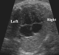
Cardiac Location
|
Apex Location |
|
||||
|
Left |
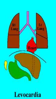
Levoposition |
Levocardia |
|
||||
|
|
Right atrium |
Right ventricle |
Cardiac apex |
Right side |
Left side |
CHD |
|
|
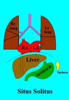
1. Situs Solitus |
Right |
Left |
Left |
Trilobed lung Liver Gallbladder IVC |
Bilobed ling Stomach Aorta Single spleen Apex of heart |
0.6-.8% |
|
|
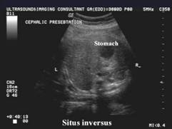
2. Situs Inversus (2%) |
Left |
Right |
Right |
Bilobed lung Stomach Single spleen Aorta Apex of heart |
Trilobed lung Liver Gallbladder IVC |
3-5% AV canal Univentricle TGV TAPVD |
|
|
3. Situs Ambiguus - asplenia |
Bilateral RA |
|
Trilobed lung Eparterial bronchi Central liver Aorta and IVC in same side of spine |
Trilobed lung Central liver Stomach in indeterminate position |
50-100% |
|
|
|
- polysplenia |
Bilateral LA |
|
Bilobed lung Midline liver Hyparterial bronchi IVC interruption with azygous continuation |
Bilobed lungs Midline liver Stomach position variable (Lt or Rt) Multiple spleens (always on same side as stomach) |
Less common than asplenia AV canal ASD PAPVD |
|
|
|
4. Visceral Heterotaxia |
Heart and / or viscera are not on one side but tend to be in the middle. |
|
|||||
|
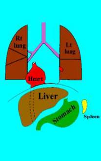
5. Mesocardia |
Heart in a midline position, stomach on left. Usually associated with a normal heart. |
|
|||||
|
|
|
|
|||||
Index
|
|
||||||
SITUS ANOMALIES |
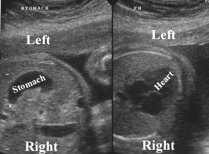
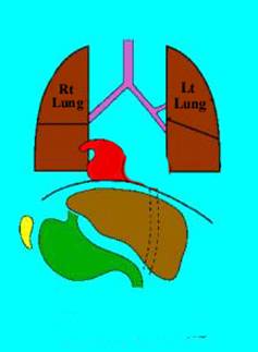
Situs refers to the position of the atria (not the cardiac apex) and abdominal visera relative to the midline. Atrial situs is determined by identification of the morphologic right or left atrial structures. The morphologic right atrium receives the heptic veins, contains the crista terminalis, and the right atrial appendage (broad based triangular shaped and contains the pectinate muscles. The left atrial appendage is long and finger-like. Typically, the morphologic left atrium is on the same side as the aortic arch (but not always). Similarly, ventricular chambers must also be identified- the left ventricle has no infundibulum so there is fibrous continuity between the mitral and aortic valves. The right ventricle is usually trapezoidal in shape and the infundibulum separates the tricuspid from the pulmonary valve. If the cardiac apex and gastric bubble are on opposite sides of the midline the patient has congenital heart disease until proven otherwise. Additionally, asplenia/ polysplenia must be ruled out.
Position of the cardiac apex:
1- Levocardia: Left sided apex (normal). Levocardia is not synonymous with situs solitus, although most patients with situs solitus have levocardia.
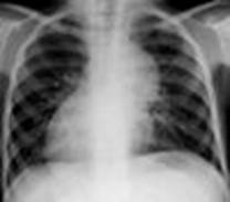
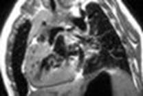
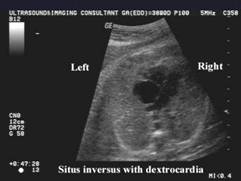
2- Dextrocardia: Cardiac apex is on the right. Most patients with situs inversus have dextrocardia.
3- Mesocardia: The bulk of the cardiac mass is in the center of the chest. There is no association with CHD.
Types of Situs:
1. Situs Solitus (
2. Situs Inversus with Mirror Image Dextrocardia: Situs inversus with dextrocardia is the usual situation. There is left-right reversal of the cardiac chambers coupled with a right sided stomach, right apex, and usually right arch. There is ventricular inversion- the left ventricle (smooth) is located on the right, and atrial inversion- right atrium is located on the left and visa versa. The right ventricle (trabeculated) remains anterior and the left atrium remains posterior- ie: there is no transposition of the ventricular or atrial chambers. Patients with Kartagener's syndrome have situs inversus with mirror image dextrocardia.
3- Situs Indeterminatus (or situs ambiguous or heterotaxia):
With true situs indeterminatus, there is abnormal arrangement of the organs and vessels. The stomach and liver are typically midline and congenital heart disease occurs in 50-100% of cases [2]. Major subcategories of situs ambiguous include- asplenia and polysplenia [2].
A. Dextroversion: Situs solitus with dextrocardia. Gastric bubble and spleen are on the left, the liver is on the right, and the cardiac apex is on the right. Generally, the aortic arch is left sided. There is NO chamber inversion- right ventricle and atrium remain on right side, and left ventricle and atrium remain on the left side; however, the normal anterior to posterior chamber relationships are lost (right ventricle becomes the posterior ventricular chamber)- ie: there is transposition of the cardiac chambers. Congenital heart disease is found in 95% of cases. Congenitally corrected transposition occurs with increased frequency in these patients.
B. Levoversion: Situs inversus with levocardia. Characterized by: Gastric bubble and spleen are on the right, and the liver on the left (situs inversus). There is typically a right arch. The majority of the cardiac mass is in the left chest, but the apex is due to the right ventricle. Chamber inversion (reversed left and right relationships): the right ventricle (atrium) is on the left and forms the cardiac apex (which will be left sided). Left ventricle (atrium) is on the right. Chamber transposition (reversed anteroposterior relationships): The right ventricle (atrium) will be the posterior ventricular (atrial) chamber. Levoversion is ALWAYS associated with congenital heart disease. Also must exclude asplenia/ polysplenia syndromes.
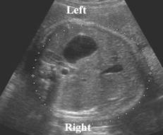
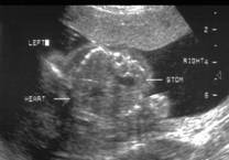
Fetal stomach / Fetal liver
|
|||
|
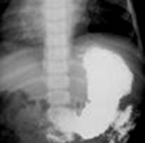
Stomach on left
/ liver on right (situs solitus) |
Stomach on right
/ liver on left (situs inversus) |
||
|
Levocardia (heart on left) |
Dextrocardia
(isolated) (normal situs) |
Levocardia
(isolated) |
Dextrocardia (with situs inversus) |
|
<1% CHD |
CHD >95% |
Almost 100% CHD |
0.3-5% CHD |
|
|
|
|
|
|
Cardiacapex on left Aortic arch on left Stomach on left IVC on right Right atrium on right Right ventricle on right Left atriun on left Left ventricle on left |
2 types:
|
Stomach + spleen on right Liver on left Right sided aortic arch (usually)
Corrected TGV DORV Systemic venous anomalies (absent IVC) Heart in left chest Right atrium on left (and is the posterior chamber) Right ventricle on left (and forms the cardiac apex) Left atrium on right Left ventricle on right |
Cardiac apex on right Aortic arch on right Stomach on right IVC on left Right atrium on left Right ventricle on left Left atrium on right Left ventricle in right VA + AV connections usually physiologic. ASD VSD AVSD |
|
|
|
|
|
Associations |
|||
|
If levorotation is present (extreme left cardiac axis
deviation >57-75 degrees) – significant risk of CHD |
Rule out
Scimitar syndrome
|
|
Rule out associated lung abnormalities especially
Kartageners syndrome |
Fetal stomach midline or left or right/ Fetal
liver midline (visceral heterotaxy)
|
|||
|
|
|||
|
Asplenia |
Polysplenia |
||
REFERENCES |
(1) Applegate KE, et al. Situs revisited: Imaging of the heterotaxy syndrome. Radiographics 1999;19: 837-852
(2) Fulcher AS, Turner MA. Abdominal manifestations of situs anomalies in adults. Radiographics 2002;22: 1439-1456