|
VENTRICULAR SEPTAL
DEFECTS |
Ventricular septal defects are the most common congenital cardiac defect, accounting for 30% of heart defects in live born infants
and 9.7% in fetuses (1,2). VSD's are associated with other cardiac malformations in about 50% of cases (3). VSD's have the
highest recurrence rate and is the most teratogen-associated defect.
ULTRASOUND
|
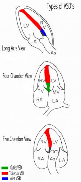
CLASSIFICATION
|
|
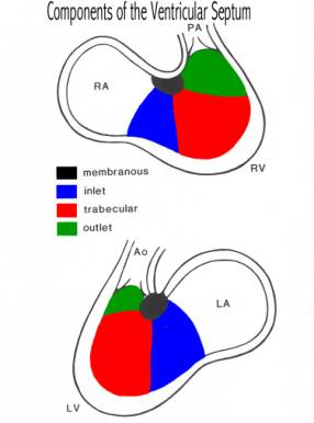
1. Membranous Defects (also called perimembranous or
infracristal). |
|
|
Perimembranous defect (80%) |
Involve the membranous septum below the aortic valve with extension to a variable degree into the adjacent portion of the septum. |
|
2. Muscular Defects (20%) - There are four subgroups
classified by the location of the defect. |
|
|
Inlet defect (5%) |
Involve the inflow tract of the right ventricle and affects the implantation of the septal chordae of the tricuspid valve They are posterior and inferior to membranous defects. |
|
Trabecular defects (5-20%) |
Occur posterior to the septal band of the crista (trabecula septomarginalis) in the midportion of the septum Often multiple, small and tortuous. |
|
Outlet defects (Conal, supracristal,subaortic or infundibular) (5%) |
Occur anterior to the septal band of the RV in the most superior portion of the interventricular septum. |
|
Apical defects (rare) |
Occur at the apex of the heart distal to the insertion of the moderator band. They may be large and difficult to identify. |
OTHER
|
Questions That Must be Answered When a VSD is
Discovered?
Table of Types of Isolated Defects Versus
Outcome at our Instititution
REFERENCES
|
- Hoffman JIE, Christianson R. Congenital heart disease in a cohort of 19,502 births with long term follow-up. Am J Cardiol 1978;42:641-647.
- Ferencz C, Rubin JD, McCarter RJ et.al. Cardiac and non-cardiac malformations: observations in a population based study. Tetralogy 1987;35:367-378.
- Goor DA, Lillehei CW. Congenital malformations of the heart. New York: Grune and Stratton 1975.