|
DOUBLE OUTLET RIGHT
VENTRICLE (DORV) |
DORV occurs when more than 50% of both the aorta and pulmonary artery arises
from the right ventricle (1), secondary to maldevelopment of the conotruncus.
CLASSIFICATION |
- DORV with a subaortic VSD (68%).
- DORV with a subpulmonary VSD (22%).
- DORV with both a subaortic and subpulmonic VSD (3%).
- DORV with a remote VSD (7%).
DORV + intact ventricular septum has been described but is very rare.
TYPES OF RELATIONSHIPS BETWEEN GREAT VESSELS |
- Aorta is posterior to and to the right of the pulmonary artery.
- Aorta and pulmonary artery are parallel, with the aorta to the right (Taussig-Bing). Aorta posterior to pulmonary artery.
- Aorta and pulmonary are parallel, with the aorta anterior and to the left (levo-malposition).
- Aorta is right and anterior to the pulmonary artery (dextra-malposition)
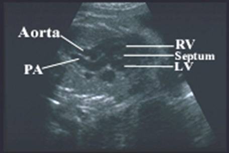
ULTRASOUND |
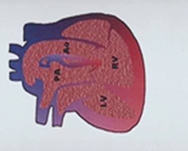
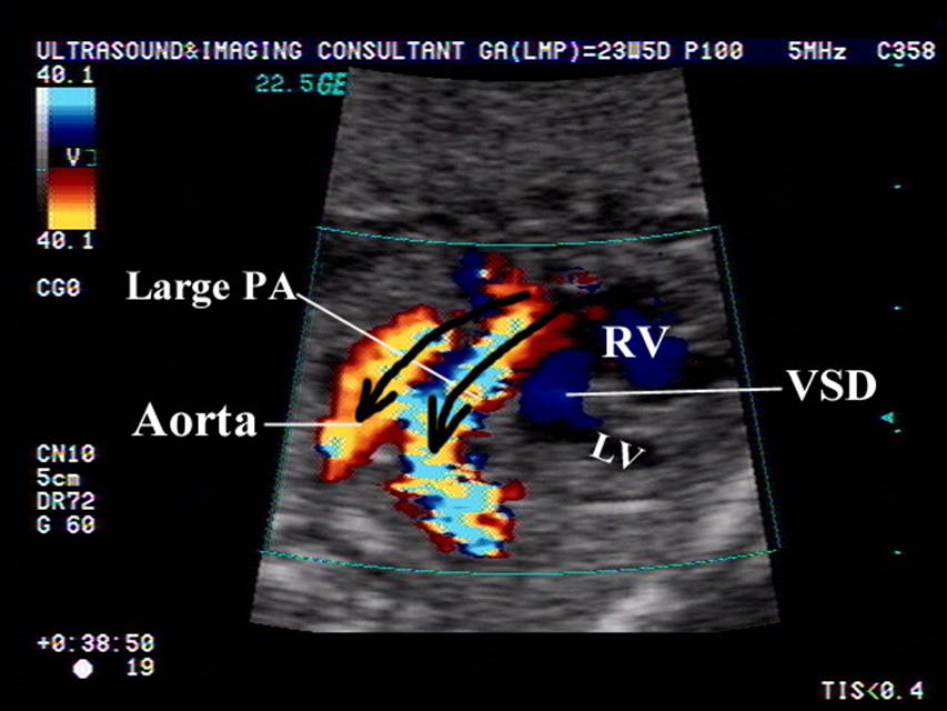
- Aorta and Pulmonary arteries arise from RV (Parallel Great Vessels).
- Aorta overriding the ventricular septum.
- Aorta posterior / parallel / anterior to pulmonary artery.
- VSD (100%).
- Pulmonary stenosis (50%).
|
|
|
|
Aorta
and PA both arise from the RV. The
aorta and PA are parallel to one another. Other
findings include: hypoplastic |
|
|
|
|
|
|
|
|
|
Video clip of Double Outlet Right Ventricle (DORV)
|
|
|
|
|
|

DIFFERENTIAL DIAGNOSIS |
- Transposition of the great vessels.
- Tetralogy of Fallot.
ASSOCIATED CONDITIONS AND SYNDROMES |
Link to Associated Conditions
and Syndromes (1)
HEMODYNAMICS |
The hemodynamics are dependent on the type of DORV and the associated anomalies.
It does not cause cardiac failure.
REFERENCES |
- Stewart PA, Wladimiroff JW, Becker AE. Early prenatal detection of double outlet right ventricle by echocardiography. Br Heart J 1985;54:340-342.