|
COARCTATION OF THE
AORTA |
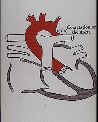
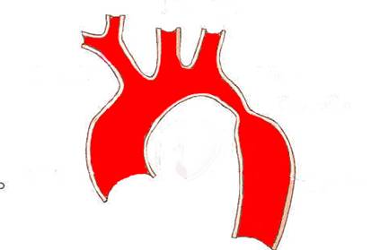
Coarctation is narrowing of the aortic lumen, usually at the ductus arteriosus. The severity of this defect ranges from slight narrowing of the distal end to severe hypoplasia of the entire arch.
TYPES OF COARCTATION |
CLASSIFICATION |
HEMODYNAMICS |
No significant change because only about 10% of the cardiac output flows
through the aortic isthmus. The descending aorta is mainly supplied via the
ductus arteriosus. The hemodynamic
effect may be greater if the aortic arch is hypoplastic.
ULTRASOUND |
- Actual area of narrowing may be difficult to identify antenatally. This may be due to normal patency of the ductus arteriosus in utero. Prenatal diagnosis usually relies on the presence of ancillary findings.
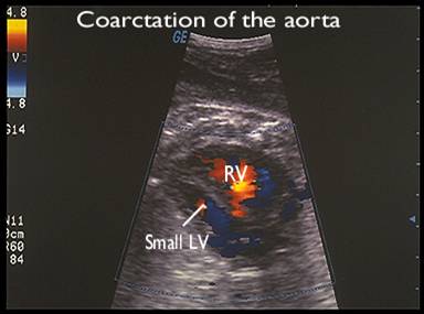
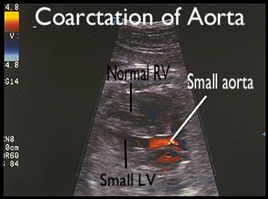
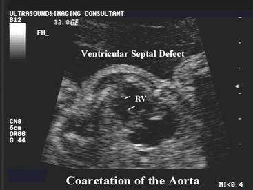
- Large RV (with a ratio over the left ventricle greater than 1.3) (3,4). Right ventricular hypertrophy has been reported.
- Small LV is a useful
sign (4,5), however some fetuses do not have
ventricular size discrepancy and some fetuses with ventricular discrepancy
do not have a cardiac lesion. It's sensitivity for the diagnosis of
coarctation of the aorta is 50-60% (6,7).
False positive (8,9) - Ventricular disproportion in the third trimester due to relative increase in the RV in comparison to the
|
|
|
|
|
|
|
|
|

|
|
Video clip of
Coarctation of Aorta - small left ventricle Video clip of
Coarctation of the Aorta – Sagittal views Video clip of
Coarctation of the Aorta – short axis
|
|
|
|
|
|

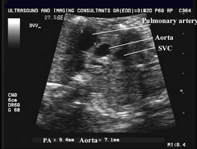
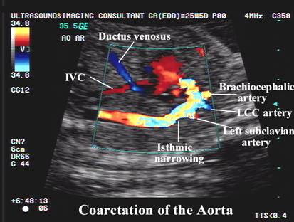
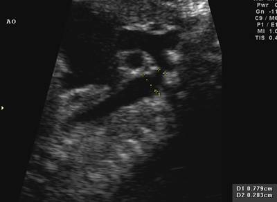
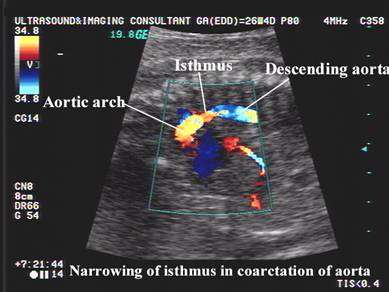
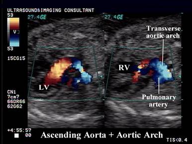
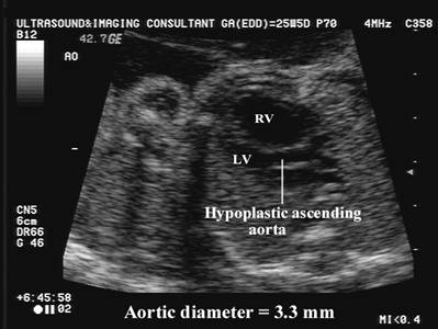
- Aortic arch - Hypoplasia of the aortic arch affects the proximal arch, most commonly between the left common carotid artery and the left subclavian artery or the isthmus, and may extend into the brachiocephalic vessels. Sonographically a small aorta at the level of the valve is present in most fetuses due to hypoplasia of the isthmus and transverse arch. Sagittal view of the arch may be normal. Hypoplasia of the transverse arch and isthmus may be detected in 80-100% of cases when adequate images of the distal arch can be obtained (14). 12/15 fetuses had a transverse arch diameter less the 3rd percentile. 10/10 fetuses had Isthmic hypoplasia with a diameter less than the third percentile. 15/18 fetuses with adequate visualization of the ascending aorta, the diameter was less than the third percentile for gestational age.
- Normograms for aortic size have been reported.
|
|
|
5.
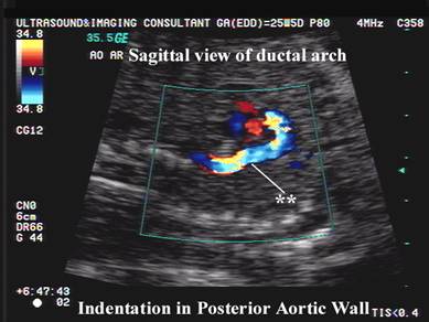
Sagittal view.
|
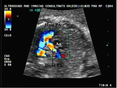
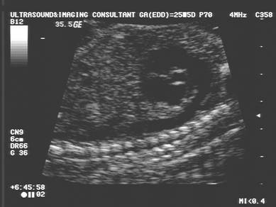
Case 1 |
|
|
|
|
|
|
|
|
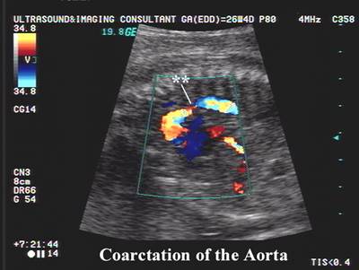
Case 2 |
|
|
|
|
|
|
|

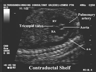
- Echogenic contralateral (contraductal)
shelf in the aortic lumen or generalized narrowing (3,4). This appears to be the least frequent finding that
can be detected on antenatal scans. Hutchins (15) described it as an
enfolded obstructive curtain in the posterior wall of the aorta, which
could represent a branch point in the aorta, resulting from increased
pulmonary artery and ductal flow relative to
aortic flow in utero.
|
|
|
|
|
|
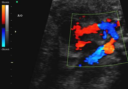
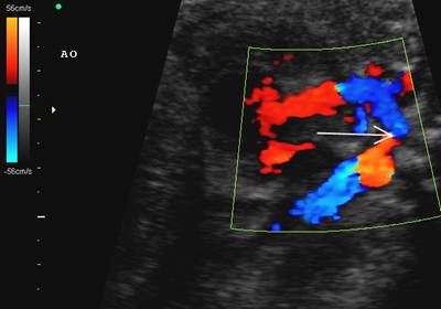
- Color Doppler may demonstrate:
- Normal flow in the aorta with normal velocities.
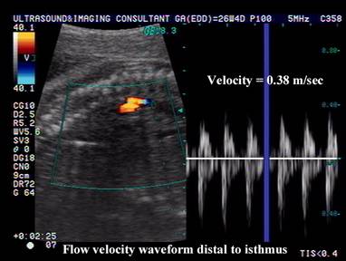
- Increased or decreased velocities distal to the coarctation (3).
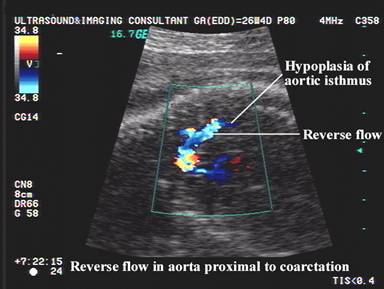
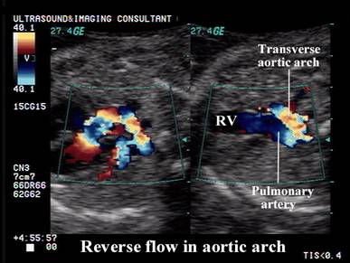
- Retrograde flow proximal to the coarctation (14).
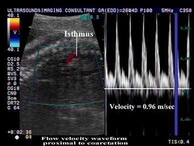
- High velocity jet that is present within the narrowed segment or just proximal to it (17).
- Turbulent flow.
Velocity change across the coarctation on pulsed doppler
|
|
|
|
|
|
|
|
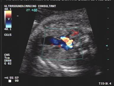
Reverse blood flow proximal to the coarctation
|
|
|
|
|
|
|
|
|
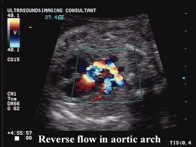
Reverse flow in the aortic arch |
Normal flow in the aortic arch |
- Color and pulsed doppler may demonstrate left to right flow through the foramen ovale (9).
- Increased flow across the tricuspid valve when compared to the mitral valve (3).
- Left axis deviation (>57°).
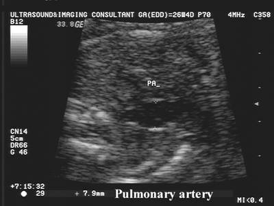
- Hornberger et.al. 1994 (14) studied prenatal scans on 20 fetuses with coarctation and 92 normal fetuses (gestational ages 18-36 wks):
- Statistically
significant difference between
- In 12 fetuses with
coarctation the aorta was significantly smaller than the pulmonary
artery. Pulmonary artery to ascending aorta ratio was 1.61 ± 0.35 when a coarctation was present
(Normally = 1.18 ± 0.06). This
suggests that aortic coarctation may be the result of diminished flow
across the aortic isthmus. Arterial growth is thought to be related to
blood flow, and distal arch hypoplasia occurs
secondary to decreased aortic blood flow relative to flow through the
pulmonary artery and ductus in utero. Decreased
- Ratio of internal
diameter of left common carotid artery to transverse arch = 0.78 ± 0.13 (
|
|
|
ASSOCIATED ANOMALIES |
- Abnormal aortic valve - bicuspid aortic valve (25-50%) or stenosed.
- Mitral valve is abnormal in 25-50% of cases (10).
- Complete heart block may coexist (11).
- Other cardiac malformations.
- PDA (33%).
- VSD (15%), ASD.
- Aortic stenosis or insufficiency.
- Truncus arteriosus, double outlet right ventricle.
- Single ventricle.
|
|
- Turners Syndrome (13-15%).
- Intracranial aneurysms.
- Diaphragmatic hernia (12).
- Situs anomalies, short umbilical cord, renal agenesis, polycystic disease of the kidney and tracheo-esophageal fistulae have been reported.
DIFFERENTIAL DIAGNOSIS |
- Small
- Relatively small
- Relatively large RV (pulmonary atresia, stenosis or regurgitation).
- Interrupted aortic arch.
- Anomalous pulmonary venous connection.
- Severe IUGR (may have ventricular disproportion) (13).
REFERENCES |
- Allan LD,
- Ferencz C, Rubin JD, McCarter RJ et.al. Cardiac and non-cardiac malformations: Observations in a population based study. Tetralogy 1987;35:367-378.
- Allan LD,
- Benacerraf BR, Saltzman DH, Sanders SP. Sonographic sign suggesting the prenatal diagnosis of coarctation of the aorta. J Ultrasound Med 1989;8:65-69.
- Emerson D, Cartier M, DiSessa T et.al. Prenatal sonographic identification of coarctation of the aorta. J Ultrasound Med 1988;7:S271.
- Kirk JS, Comstock CH, Lee W et.al. Sonographic screening to detect fetal cardiac anomalies: A 5-year experience with 111 abnormal cases. Obstet Gynecol 1997;89:227-232.
- Brown DL. Sonographic assessment of fetal arrhythmias. AJR 1997;169:1029-1033.
- Brown DL, Durfee SM, Hornberger LK. Ventricular discrepancy as a sonographic sign of coarctation of the fetal aorta: How reliable is it? J Ultrasound Med 1997;16:95-99.
- Sharland GK, Chan K, Allan LD. Coarctation of the aorta: Difficulties in prenatal diagnosis. Br Heart J 1994;71:70-75.
- Becker AE, Becker MJ, Edwards JE. Anomalies associated with coarctation of the aorta. Circulation 1979;41:1067-1072.
- Machado MV, Tynan MJ, Curry PV et.al. Fetal complete heart block. Br Heart J 1988;60:512-515.
- Siebert JR, Hass JE, Beckwith JB. Left ventricular hypoplasia in congenital diaphragmatic hernia. J Pediatr Surg 1984;19:567-571.
- Siassi B. Normal and abnormal transitional circulation in the IUGR infant. Semin Perinatol 1988;12:80-83.
- Hornberger LK, Sahn DJ, Kleinman CS et.al. Antenatal diagnosis of coarctation of the aorta: A multicenter experience. J Am Coll Cardiol 1994;23(2):417-423.
- Hutchins GM. Coarctation of the aorta explained as a branch pint of the ductus arteriosus. Am J Pathol 1971;63:203-209.
- Rudolph AM, Heymann MA, Spitznas U. Hemodynamic considerations in the development of narrowing of the aorta. Am J Cardiol 1972;30:514-525.
- Snider RA, Serwer
GA. Abnormal vascular connections and structures: In: Snider RA,