|
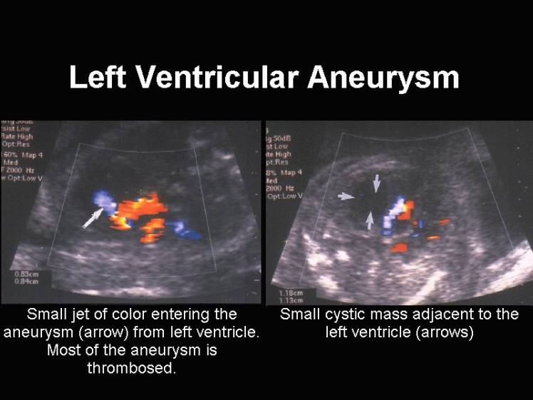
LEFT VENTRICULAR
ANEURYSM |
Congenital left ventricular aneurysm is a rare cardiac anomaly that has been detected antenatally (1-7). A ventricular aneurysm is an outpouching of the fibrous ventricular wall in conjunction with akinesis and dyskinesis. The aneurysm wall contains myocardium and fibrous tissue. The neck of the defect is usually broad, and the defect may be uni- or multilobulated. These defects are usually not associated with other intracardiac anomalies. The cause of a congenital cardiac aneurysm id obscure, however prenatal infection, trauma and ischemia of the myocardium could be implicated.
A left ventricular diverticulum is a fingerlike projection communicating with the left ventricular apex. It has a smooth non-contractile wall, and involved the apex of the heart. The neck of the defect has a well-developed muscular wall and is associated with anomalies of midline structures of the body such as sternum, pericardium, heart (ASD, VSD), diaphragm and anterior abdominal wall (8-11).
A false aneurysm or pseudoaneurysm is a localized myocardial rupture limited
by adherent pericardium and fibrous tissue (11).
|
Classification |
Etiology |
||
|
- Subaortic |
- Unknown? |
||
|
|
|||
|
Reference |
GA at diagnosis |
Other Findings |
Outcome |
|
(1) |
32 weeks |
Cardiac arrhythmia |
Delivery at 41
weeks |
|
(2) |
35.5 weeks |
No cardiac failure |
Delivery at 39
weeks |
|
(3) |
29 weeks |
Cardiac failure |
Intrauterine demise |
|
(4) |
24 weeks |
Pericardial
effusion |
Intrauterine
demise |
|
(5) |
28 wks |
No cardiac failure
or arrhythmia |
Demise post
surgical repair |
|
(7) |
21 wks |
Ascites, pleural
effusions and subcutaneous edema at 27 wks. |
Intrauterine
demise at 27 weeks. |
|
|
Video clip of a Left Ventricle
Diverticulum
|
|
|
|
|
|

DIFFERENTIAL DIAGNOSIS |
- Ventricular diverticulum (associated with defects in the diaphragm, pericardium and abdominal wall) (6).
- Developmental defect of the septum transversum.
COMPLICATIONS |
Prenatal – spontaneous rupture, arrhythmia (ventriculat tachycardia), congestive heart failure, and mass effect on the adjacent lung with associated underdevelopment.
Postnatal – thromoboembolism, bacterial endocarditis, angina from compression of coronary arteries by the expanding aneurysm (12).
REFERENCES |
- Gembruch U, Steil E, Redel DA et.al. Prenatal diagnosis of left ventricular aneurysm. Prenat Diagn 1990;10:203-209.
- Jacobson RL, Perez A, Meyer R et.al. Prenatal diagnosis of fetal left ventricular aneurysm: a case report and review. Obstet Gynecol 1991;78:525-527.
- Goncalves LF, Simms J, Jeanty P. Aneurysm, left ventricle. Fetus 1992;1:7-10.
- Sherman SJ, Leenhouts KH, Utter GO et.al. Prenatal diagnosis of left ventricular aneurysm in the late second trimester: a case report. Ultrasound Obstet Gynecol 1996;7:456-457.
- Suchet IB Unpublished case.
- Singh A, Katov H, Zavoral J et.al. Congenital aneurysms of the left ventricle. Am Heart J 1980;99:25-32.
- Chaubal N, Dighe M, Shah M et.al. Congenital left ventricular aneurysm. J Ultrasound Med 2004;23:125-128.
- Hornberger LK, Dalvi B, Benacerraf BR. Prenatal sonographic detection of cardiac aneurysms and diverticuli. J Ultrasound Med 1994;13:967-970
- Triestman B, Cooley DA, Lufschanowski R, Leachman RD. Diverticulum or aneurysm of left ventricle. Am J Cardiol 1973;32:119-123.
- Crawford FA. Left ventricular aneurysm. Ann Thorac Surg 1991;52:173-174.
- Jacobson RL, Perez A, Meyer RA et.al. Prenatal diagnosis of left ventricular aneurysm: a case report and review. Obstet Gynecol 1991;78:525-528.
- Papagiannis J, Van Pragh R, Schwint O et.al. Congenital left ventricular aneurysm, clinical imaging, pathologic and surgical findings in 7 new cases. Am Heart J 2001;141:491-499.