|
HYPERTROPHIC
(OBSTRUCTIVE) CARDIOMYOPATHY |
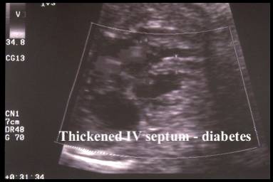
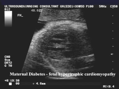
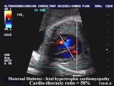
Infants of diabetic mother are at risk of developing hypertrophic
cardiomyopathy (1). 35-75% have transient hypertrophic cardiomyopathy.
ULTRASOUND |
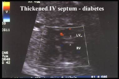
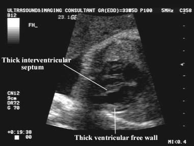
- Thickening of the interventricular septum (2,3).
- Thickening of the ventricular
free walls (although the thickened wall may be evident by 20 weeks of
gestation it is mainly evident during the late second trimester) (2,3).
The increase of cardiac wall thickness has been shown to influence fetal
cardiac motion (4,5).
At birth, the hypertrophic changes of the myocardium regress to normal over a period of months and are usually no longer present at one year of age (6). - Systolic and diastolic dysfunction of the neonatal heart, which may result in cardiac failure in the immediate postnatal period (6).
- These changes have been described antenatally and have been shown to progress with fetal growth.
- The increased cardiac size does not reflect the macrosomia that is present in fetuses of diabetic mothers, but represents a selective organomegaly (2,4).
- Doppler Assessment
- E/A values are low
- Impaired development of ventricular compliance in the fetuses secondary to cardiac wall thickening (5).
- Polycythemia is frequently present at birth in infants of diabetic mothers. This increases blood viscosity which may reduce preload and affect the E/A ratio (7).
- Peak velocities at the level of the aortic and pulmonary outflow tracts are significantly higher in fetuses of diabetic mothers. This may be due to reduced outflow tract dimensions, decreased afterload increased cardiac contractility or increased flow volume Increased intracardiac flow volume secondary to a relatively large fetal size since cardiac output is a function of fetal weight (4).
- The percentage of reversed flow in the IVC is increased (if the reverse is above 2 SD's from the expected mean for gestation, a lower pH in the umbilical artery is present) (8).
|
|
|
|
|
|
|
|
|
REFERENCES |
- Gutgesell HP, Speer ME, Rosenberg HS. Characterization of cardiomyopathy in infants of diabetic mothers. Circulation 1980;61:441-450.
- Rizzo G, Arduini D, Romanini C. Cardiac function in fetuses of type I diabetic mothers. Am J Obstet Gynecol 1991;164:837-843.
- Vielle JC, Sivekoff M, Hanson R et.al. Interventricular septal thickness in fetuses of diabetic mothers. Obstet Gynecol 1992;79:51-54.
- Weber HS, Copel JA, Reece A et.al. Cardiac growth in fetuses of diabetic mothers with good metabolic control. J Pediatr 1991;118:103-107.
- Rizzo G, Arduini D, Romanini C. Accelerated cardiac growth and abnormal cardiac flows in fetuses of type I diabetic mothers. Obstet Gynecol 1992;80:369-376.
- Reller MD, Kaplan S. Hypertrophic cardiomyopathy in infants of diabetic mothers: an update. Am J Perinatol 1988;5:353-358.
- Widness J, Susa J, Garcia J. Increased erythropoiesis and elevated erythropoietin levels in infants born to diabetic mothers and in hyperinsulinemic rhesus fetuses. J Clin Invest 1981;67:637-641.
- Rizzo G, Capponi A, Rinaldo D et.al. Inferior cava velocity waveforms predict neonatal complications in fetuses of insulin dependent diabetic mothers. J Maternal Fetal Invest 1994;4: