|
TRICUSPID VALVE
DYSPLASIA |
Tricuspid valve dysplasia, absence of the tricuspid valve leaflets, Uhl's
and Ebstein's anomaly are a spectrum of diseases characterized by dysplasia,
hypoplasia or aplasia of the tricuspid valve or right ventricular myocardium or
both.
|
Case
1 |
|
|
|
|
|
|
|
|
|
|
|
Case
2 |
|
|
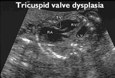
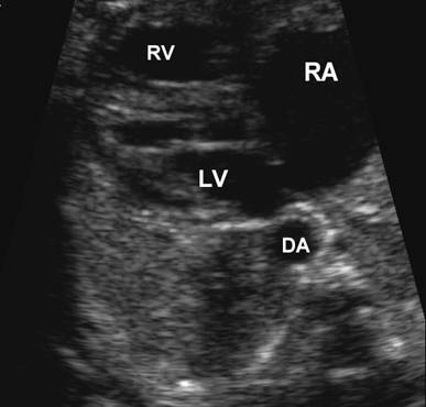
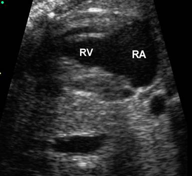
Subcostal view – large right
atrium (RA), normal right ventricle (RV) and left ventricle ( |
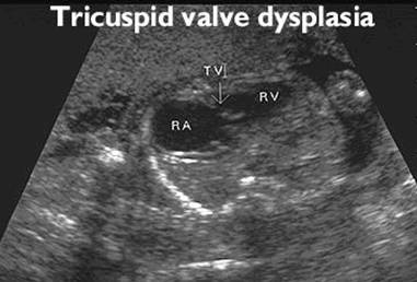
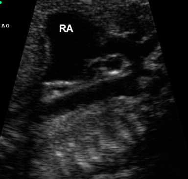
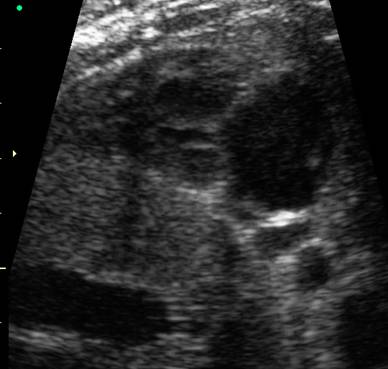
Sagittal view – large right atrium |
|
|
|
|
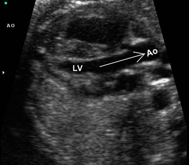
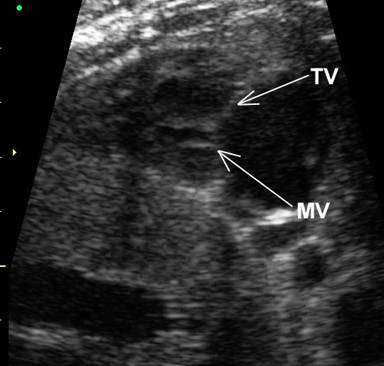
Normal position of mitral and tricuspid
valves. This excludes Ebstein’s anomaly as the cause of RA enlargement |
Normal |
|
|
|
|
Large
RA and normal size RV |
|
|
|
|
|
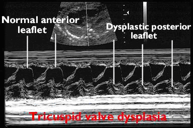
Mitral and tricuspid valves at normal
levels (no apical displacement of tricuspid valve) |
|
|
|
|
|
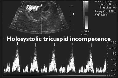
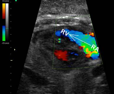
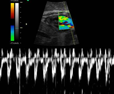
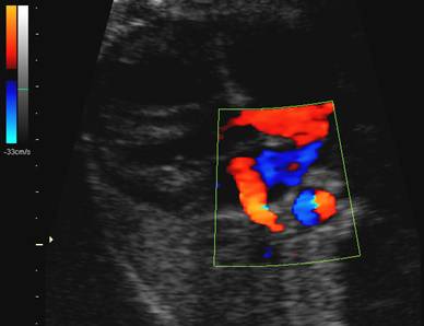
Marked
tricuspid regurgitation due to valvular dysplasia |
|
|
|
|
|
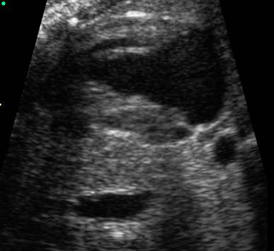
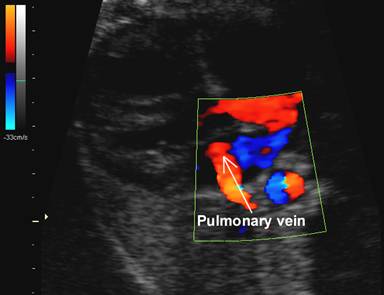
Normal
pulmonary vein draining into LA. This excludes total anomalous pulmonary
venous return |
|
|
|
|


|
|
Video clip of Tricuspid Valve Dysplasia
|

DIFFERENTIAL DIAGNOSIS |
|
Tricuspid valve dysplasia |
Valve leaflets are thickened and may be redundant or
hypoplastic. Left cardiac axis deviation. |
|
Attachment of the septal and posterior leaflets are
displaced to the right |
|
|
No tricuspid valve leaflets are visible. |
|
|
Normally formed and attached leaflets |
|
|
|
REFERENCES |
- Hornberg LK, Sahn DJ, Kleinman CS et.al. Tricuspid valve disease with significant tricuspid insufficiency in the fetus: Diagnosis and outcome. J Am Coll Cardiol 1991;17:167.
- Benson CB, Brown DL, Roberts DJ. Uhl's anomaly of the heart mimicking Ebstein's anomaly in utero. J Ultrasound Med 1995;14:781-783.