|
ANEYRYSM OF THE ATRIAL SEPTUM |
Redundant septum primum flap, also known as Foramen Ovale Aneurysm, Atrial Septal Aneurysm (ASA), or Aneurysm of Septum Primum.
An atrial septum aneurysm is defined as dilatation of the atrial septum with bulging of the septum at least half the distance to the left atrial wall.
The prevalence of ASA in the general population is unknown as it may be easily overlooked or ignored on routine fetal echocardiography.
Reported frequency: 60-588:10,000 (1,2), 7.6% (3), 0.38% (4).; 0.6-1.7% of fetuses referred for echocardiographic examination (5,6).
EMBRYOLOGY |
· The thin membranous septum primum divides the fetal atria early in embryonic development.
· The thicker septum secundum grows along the septum primum, and contains the foramen ovale.
· The foramen ovale allows normal right-to-left atrial shunting during fetal life.
· Flow across the foramen ovale displaces the septum primum into the left atrium.
· The septum primum is relatively pocket-shaped and is not intact along the entire extent of the inter-atrial septal wall, allowing blood to pass from right to left atrium.
· Because the opening of the septum primum is not directly over with the foramen ovale, the septum primum will effectively close the foramen ovale in early neonatal life as left atrial pressure exceeds right atrial pressure and the septum primum adheres to the septum secundum.
The origin of ASA is unknown but it has been considered as a congenital variant perhaps due to weaker septum primum tissue.2 Observations in the neonatal group however suggest that at least some redundancies may be acquired. This hypothesis was based on the fact that atrial septal aneurysms tend to get worse in patients with abnormal atrial hemodynamics, but tend to resolve in patients with normal hemodynamics (7).
ULTRASOUND |
This is primarily a defect of septum primum which results in:
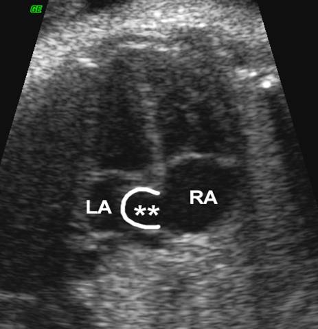
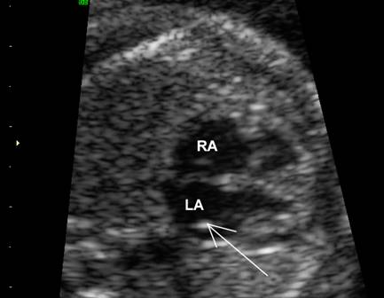
- Bulge of septum rimum that exceeds half the diameter of the LA chamber on the four-chamber view.
- Loss of the normal biphasic motion of foramen ovale.
- Cardiac arrhythmias.
The size of the FO ranged from 5.8-11.2 mm, with Z values for gestational age ranging from - 0.2 to + 5.5 (mean, 1.9).
The FO was abnormally large in 2 with Z values 5.7 and 4.2, and within normal limits in the other 7.
The maximum excursion of the flap was 51%-74% (mean, 65%) of the left atrial width. The neonatal echocardiogram showed an ASA in 4/6.
A secundum ASD, 4-5 mm in diameter, was found in the short-term postnatal follow-up in 4/9. No arrhythmias or other congenital heart defects were noted.
|
Case
1 |
|
|
|
|
|
|
|
|
|
|
|
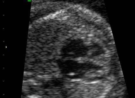
Case
2 |
|
|
|
|
|
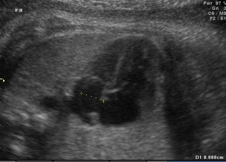
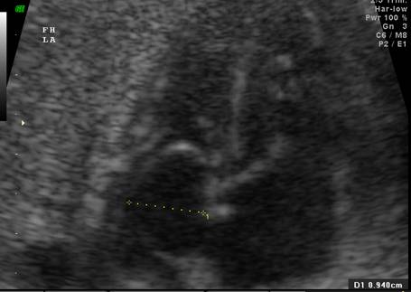
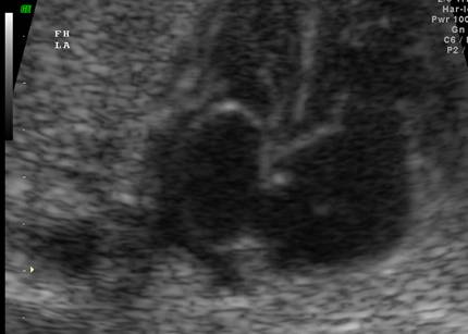
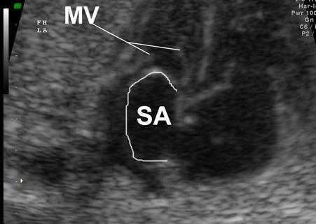
Case
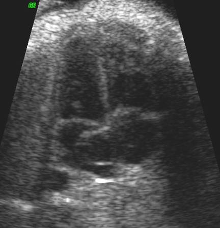
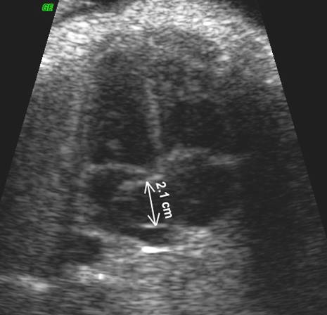
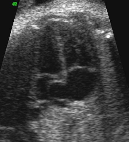
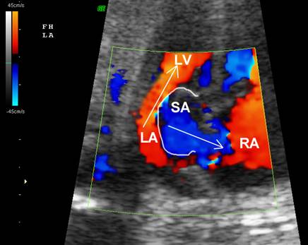
3 Large septal aneurysm extending >50%
across LA chamber Aneurysm bulging through mitral valve
(MV) No atrial septal defect. |
|
|
|
|
|
|
|
|
|
|
|
|
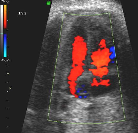
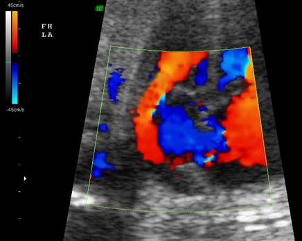
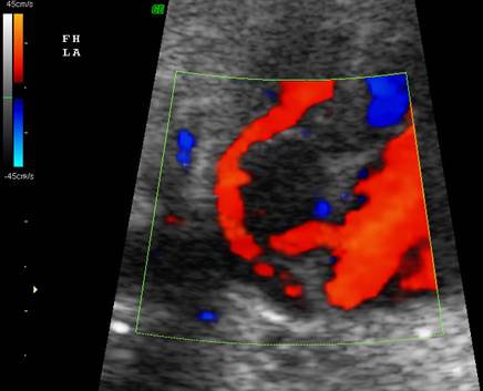
Above panels: Retrograde
flow of blood from left to right atrium. Note
the lateral displacement of blood flow from LA to Large
aneurysm protruding through the mitral valve. Botton
Panel: Normal
flow from right atrium to left atrium across the foramen ovale. |

|
|
|
|
|
|
|
|

ASSOCIATED ANOMALIES |
- Atrial septal defect.
- Tricuspid atresia.
- Hypoplastic right heart.
- Aortic stenosis.
- Transposition of the great vessels.
- Ebsteins anomaly.
- AV valve and pulmonary venous
obstruction.
DIFFERENTIAL DIAGNOSIS |
- Left atrial appendage.
- Left atrial thrombus.
- Anomalous left SVC.
PROGNOSIS |
Foramen of Ovale
Aneurysms are often Isolated .
It may be associated cardiac arrhythmia in up to 67% of cases, which
generally resolves at birth.
- The majority of these arrhythmias are solitary PAC's;
- There may rarely be short runs of - or even sustained - SVT.
A strong association exists between the presence of ASA and PACs (5,9). The mechanism is not entirely clear.
Possible explanations may include:
1) the base of the foraminal flap may cause mechanical irritation to the sino-atrial node, or
2) it may be the interaction between the foraminal flap and the left atrial wall, or
3)
ASA may cause
blocking of SA-AV node transmission
Whatever the precise cause, PACs tend to resolve spontaneously in neonatal life when the foraminal flap adheres to the septum secundum and no longer protrudes into the left atrium (8-10).
REFERENCES |
- Stewart PA, Wladimiroff J; Fetal atrial arrhythmias associated with redundancy/aneurysm of the foramen ovale. JCU 16: 643, 1988.
- Papa M, Fragasso G, Camesasca C et.al. Atrial septal aneurysm in high risk fetuses. Italian Heart J 2002;3:318-321.
- Gloeb DJ,
- Stewart PA et.al. Fetal Atrial Arrhythmias Associated with redundancy/Aneurysm of the Foramen Ovale. J. Clin Ultrasound 1988;16:643-650.
- Kachalia P et.al. In Utero Sonographic Appearance of the Atrial Septum Primum and Septum Secundum. Ultrasound Med 1991;10:423-426.
- Wolf WJ, Casta A, Sapire D: Atrial Septal Aneurysms in Infants and Children. Am Heart J 1987; 113:1149-1153
- Pinette MG, Pan Y, Pinette SG, et al: Fetal Atrial Septal Aneurysm. JRM 1997; 42: 459-462.
- Rice MJ, McDonald RW, Reller MD: Fetal Atrial Septal Aneurysm: A Cause of Fetal Atrial Arrhythmias. J Am Coll Cardiol 1988; 12:1292-7.
- Toro L, Weintraub G, Shiota T, et al: Relation Between Persistant Atrial Arrhythmias aand Redundant Septum Primum Flap (Atrial Septal Aneuyrsm) in Fetuses. Am J Cardiol 1994: 73:711-3.