|
MECONIUM ILEUS |
Meconium ileus is the most common fetal intestinal disorder occurring in
10-15% of cases (1-3). It is the third most common cause of neonatal bowel
obstruction after atresia and malrotation (1). This is because cystic fibrosis (CF)
patients have meconium containing increased protein and decreased water making
it thick and viscous (1).
ETIOLOGY |
ULTRASOUND |
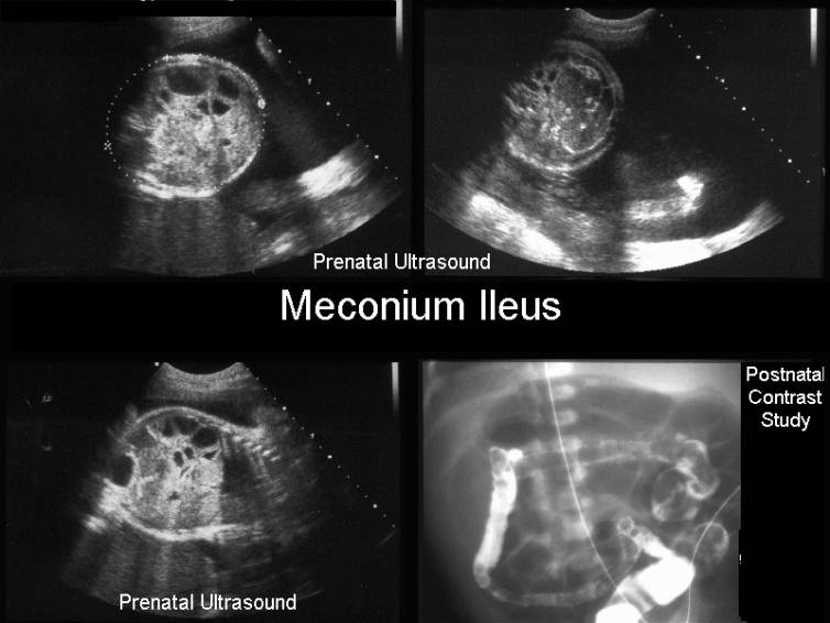
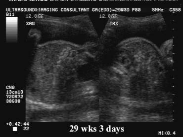
- Complex echogenic mass (small intestine dilated with meconium) in the lower abdomen (4).
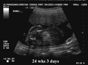
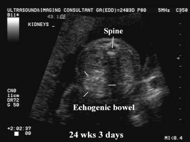
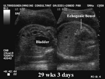
- Usually presents in the late second trimester as echogenic bowel or in the third trimester as echogenic or dilated loops of bowel. This pattern occurs because meconium begins to accumulate in the fetal bowel in the second trimester (1,3).
- Dilated fluid filled loops of bowel.
- Polyhydramnios.
- Fetal stomach may be dilated (1).
- Intraperitoneal calcifications, echogenic masses or pseudocyst may be present if meconium ileus is complicated by meconium peritonitis.
|
|
|
|
|
|
|
|
|
|
|
|
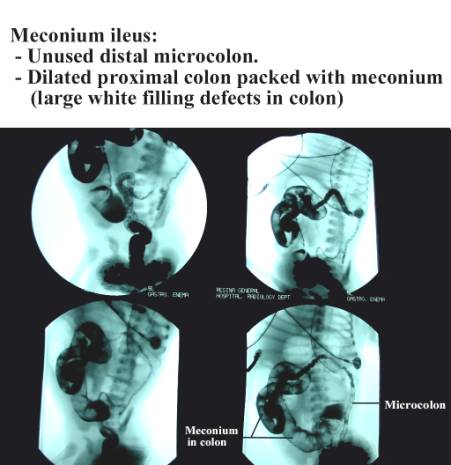
COMPLICATIONS |
- Small bowel obstruction.
- Meconium peritonitis.
REFERENCES |
- Goldstein R, Filly R, Callen P. Sonographic diagnosis of meconium ileus in utero. J Ultrasound Med 1987;6:663.
- Nyberg DR, Hastrup W, Watts H et.al. Dilated fetal bowel: A sonographic sign of cystic fibrosis. J Ultrasound Med 1987;6:257.
- Denholm TR, Harte CC, Edwards WH et.al. Prenatal sonographic appearance of meconium ileus in twins. AJR 1984;143:371.
- Grimaldi L, Ternent CA, Sheehan FR. Detection of complications of cystic fibrosis. J Ultrasound Med 1994;13:997-999.