|
HIRSCHSPRUNG’S
DISEASE |
Hirschsprung's disease is caused by the absence of the ganglion cells of the myenteric plexus extending proximally from the distal colon. This is thought to occur secondary to the arrest of the craniocaudal migration of neuroblasts. This results in a normal or reduced caliber of the aganglionic segment, and a functional obstruction with distention of the more proximal normal colon.
- 80% involve the distal 10cm of large bowel.
- 10-15% involve a longer segment of colon.
- 5-10% have total colonic involvement.
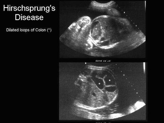
ULTRASOUND (1,2) |
- Dilated loops of colon (peripheral location, lack of peristalsis, haustral markings or pelvic location). The ultrasound finding of dilated colon does, however have a poor predictive value and in the majority of cases does not represent colonic or anal obstruction (1).
- Dilated loops of colon with obstruction is usually not detected prior to 25 weeks gestation (3,4).
- Enterolithiasis.
- Increased abdominal circumference.
- Polyhydramnios.
|
|
|
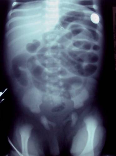
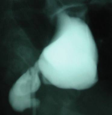
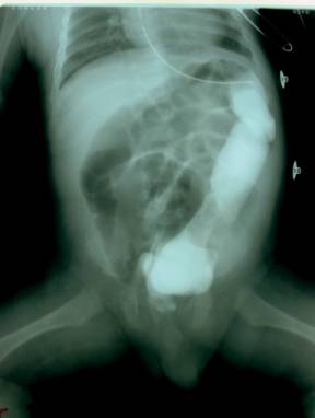
Postnatal Abdominal Radiograph and Barium Enema |
|
|
|
Dilated loops
of small and large bowel. Paucity of gas in rectum and sigmoid region. Transitional area noted at recto-sigmoid junction on barium enema with Colonic dilatation proximal to this area. |
|
|
|
ASSOCIATED ANOMALIES |
- Esophageal dysmotility syndromes.
- Down's Syndrome (3% of Down's syndrome have Hirschsprung's disease).
- Malrotation.
- Ileal and colonic atresias.
- Neurocristopathies (neuroblastoma, phaeochromocytoma, neurofibromatosis, Waardenberg's syndrome, and MEN IIA).
- Metaphyseal dysplasia of the McKusick-Kaufman type.
- Cat-eye syndrome.
- Congenital rubella.
REFERENCES |
- Vermesh M, Mayden KL, Confino E et.al. Prenatal sonographic diagnosis of Hirschsprung's disease. J Ultrasound Med 1986;5:37-39.
- Eliyahu S, Yanai N, Blondheim O et.al. Sonographic presentation of Hirschsprung's disease. A case of an entire aganglionic colon and ileum. Prenat Diagn 1994;14:1170-1172.
- Belin B, Corteville JE, Langer JC. How accurate is prenatal sonography for the diagnosis of imperforate anus and Hirschsprung's disease? Pediatr Surg Int 1995;10:30-32.
- Harris RD, Nyberg DA, Mack LA. Anorectal atresia: prenatal sonographic diagnosis. AJR 1987;149:395-400.