|
BILATERAL RENAL
AGENESIS |
Renal agenesis, the complete congenital absence of renal tissue, results from the failure of the ipsilateral ureteric bud to contact the nephrogenic blastema. This may be because of failure of ureteral bud development or an inherent deficiency in the metanephric blastema.
Bilateral renal agenesis is characterized by absence of the kidneys, ureters and renal arteries. The bladder may be absent or hypoplastic.
- 1-4 / 10,000 births (1) with a 2.5:1 male preponderance (1).
- Etiology unclear, but it is uniformly lethal.
- Pattern of inheritance and recurrence risk is complex.
- Most cases are sporadic (familial risk of recurrence = 3-5%)(2).
- Seen in some chromosomal disorders, autosomal dominant or recessive syndromes, with non genetic and sporadic syndromes (3).
- Risk: Previous family history (13%), previous affected sibling (3%) and with two previous affected siblings (30%) (4,5)
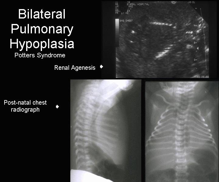
- Infants with absent kidneys have clinical features called Potter's sequence.
· It is essential to make the correct diagnosis as the mortality rate is 100%, and will avoid the necessity of performing an emergency cesarean section (severe IUGR or premature rupture of membranes may have a similar appearance.
· Severe oligohydramnios (may only be seen after 16 weeks as there are many non renal sources until this time).
· Non-visualization of the fetal bladder.
· Absent fetal kidneys. This is often difficult to visualize in the presence of oligohydramnios.
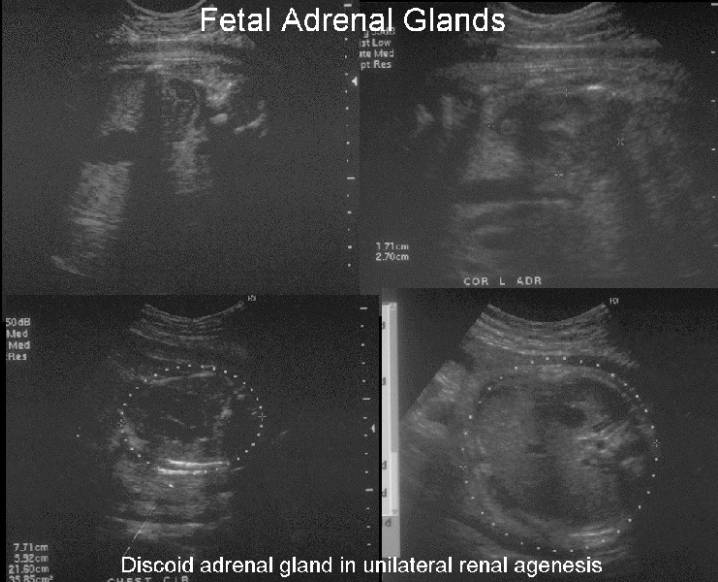
· The adrenal glands may also assume an elongated reniform or discoid shape and be confused with normal kidneys (6). According to Dunnick, in 8% to 10% of cases there is ipsilateral adrenal gland absence despite apparently unrelated embryologic development (8).
· Renal agenesis.
o The adrenals assume a reniform shape making the early diagnosis of renal agenesis difficult.
o Adrenals may completely fill the renal fossa.
o The adrenal glands may appear more globular and simulate small but present kidneys. Autopsy studies have shown this phenomenon to be caused by a change in the normal-sized adrenal’s shape, rather than adrenal hypertrophy (9). Looking at orthogonal images may avoid this problem (10).
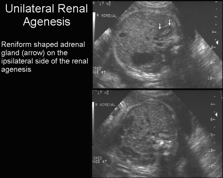
o Hoffman et al (11) reported a flat or "lying down" adrenal in 48% of 23 fetuses and 6 neonates retrospectively studied by US because of apparent renal agenesis or ectopia and suggested that the normal shape of the adrenal gland which usually "caps" the kidneys is affected by presence of an ipsilateral kidney.
o
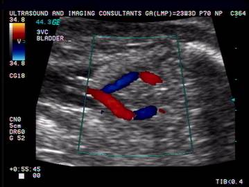
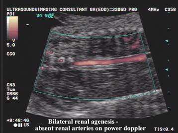
· Color doppler of the renal arteries demonstrate their absence (7).
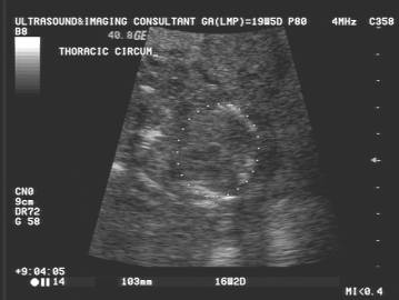
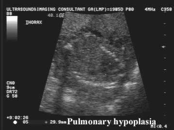
· Bell-shaped thorax (pulmonary hypoplasia) - mid / late 3rd trimester.
· Compression deformities of the extremities (clubfoot, flexion contractures, joint dislocations).
Bilateral
Renal Agenesis
·
Absent fetal bladder between the umbilical arteries. ·
Anhydramnios. |
|
|
|
|
|
|
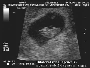
Bilateral
Renal Agenesis Normal
amniotic fluid at 8 wks 3 days Anhydramnios at 19 weeks |
|
|
|
|
|
|
|
|
|
|
|
|
|
|
|
ASSOCIATED NON GENITOURINARY ANOMALIES |
Link to Non genitourinary anomalies associated with
bilateral renal agenesis.
Differentiating
Bilateral Renal Agenesis From Sirenomelia
Syndromes
In Which Bilateral Renal Agenesis is a Component
REFERENCES |
- Carter CO, Evans K. Birth frequency of bilateral renal agenesis. J Med Genet 1981;18:158.
- Carter CO. The genetics of urinary tract malformations. J Genet Hum 1984;32:23.
- Romero P, Pilu G, Jeanty P et.al. Prenatal diagnosis of congenital anomalies.
- Carter CO, Evans K, Pescia G. Family study of bilateral renal agenesis. J Med Genet 1979;16:176.
- Roodhooft AM, Birnholz JC, Holmes LB. Familial nature of congenital absence and severe dysgenesis of both kidneys. N Engl J Med 1984;310:1341.
- McGahan JP, Myracle MR. Adrenal hypertrophy: possible pitfalls in the sonographic diagnosis of renal agenesis. J Ultrasound Med 1986;5:265.
- DeVore GR. The value of color doppler sonography in the diagnosis of renal agenesis. J Ultrasound Med 1995;14:443-449.
- N.R. Dunnick,
C. Sandler, E.S. Amis,
Jr et al.. Textbook of Uroradiology ((ed 2).),
Williams & Wilkins,
- P. Dubbins, A. Kurt, R. Wapner et al., Renal agenesis: spectrum of in utero findings. J Clin Ultrasound 9 (1981), pp. 189–193.
- S. Droste, J. Fitzsimmons, J. Pascoe-Mason et al., Size of the fetal adrenal in bilateral renal agenesis. Obstet Gynecol 76 (1990), pp. 206–209.
- C.K. Hoffman, R.A. Filly and P.W. Callen, The "lying down" adrenal sign: A sonographic indicator of renal agenesis or ectopia in fetuses and neonates. J Ultrasound Med 11 (1992), pp. 533–536.