|
URETEROPELVIC JUNCTION OBSTRUCTION (UPJ) |
UPJ obstruction is the most common congenital malformation of the urinary tract and the most common cause of neonatal and fetal hydronephrosis (1).
PUV occurs in 1 in every 5,000 to 8,000 boys; their most common cause of
urinary tract obstruction
ETIOLOGY |
- Most cases appear to represent functional rather than fixed lesions, with disordered smooth muscle arrangement in the UPJ impeding formation and propulsion of the urine bolus (2).
- Obstruction is caused by redundant membranous folds found within the posterior urethra causing variable degrees of obstruction.
- In a minority of cases, fibrous adhesions, kinks, ureteral valves and aberrant vessels may be responsible for the stenosis (3).
CLASSIFICATION |
Link to
Classification of Hydronephrosis
ULTRASOUND |
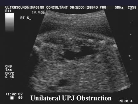
- Unilateral (70%) (4).
|
|
|
- Bilateral (30%).
- Involvement is usually asymmetric.
- Severe obstruction rarely occurs.
- Milder forms are unlikely to be fatal (5).
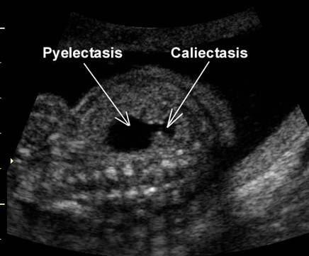
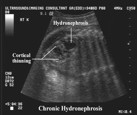
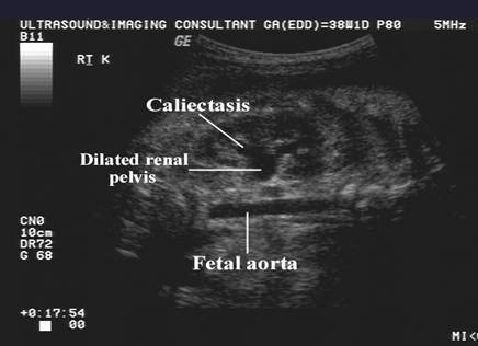
- Variable caliectasis to marked pyelectasis and thinning of the renal cortex in severe chronic obstruction (may appear as a unilocular cystic mass). Irreversible parenchymal damage is unusual.
|
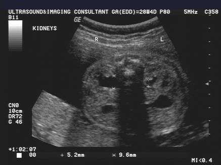
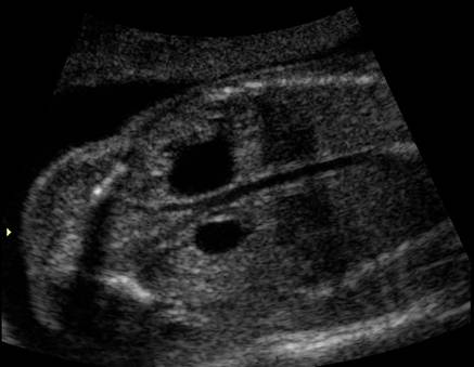
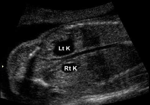
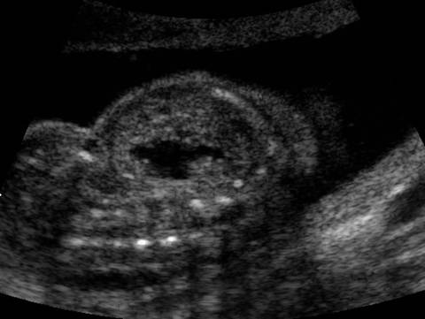
Bilateral (left greater than right)
pyelectasis and calicetasis at 20 weeks of gestation |
|
|
|
|
|
|
|
|
|
|
|
|
|
|
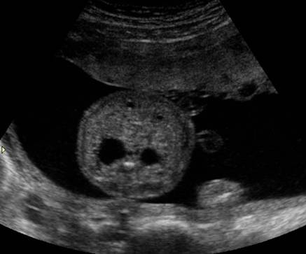
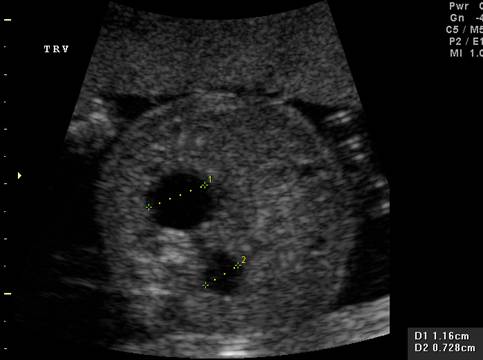
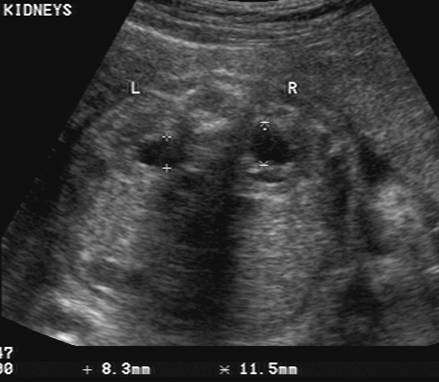
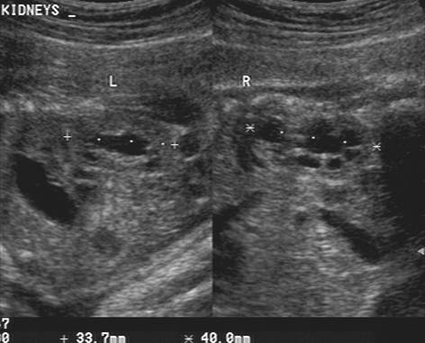
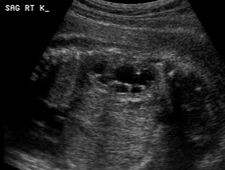
Case 2 |
|
|
|
|
|
|
|
|
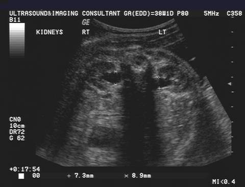
Bilateral hydronephrosis
and market cortical thinning on both kidneys |
|
|
|
|
|
|
|
- Significant abnormalities of the ureters or bladder are not normally present. Normal bladder emptying should be present.
- Amniotic fluid is usually normal unless there is severe disease of the contralateral kidney.
- Degree of hydronephrosis depends on the onset, duration and degree of obstruction that is present>
- Early onset (8-10 weeks) will probably result in a multicystic dysplastic kidney.
- Late onset results in variable hydronephrosis but without dysplastic renal parenchymal changes.
- Dilatation may be intrarenal (involves calyces and renal pelvis) or extrarenal (involves the extrarenal
portion of the renal pelvis).
Intrarenal hydronephrosis is involved with more renal parenchymal damage (6).
ASSOCIATED ANOMALIES |
- Renal anomalies are seen in up to 27% of cases (7). They include vesicoureteric reflux, lower ureteral obstruction, contralateral renal agenesis, meatal stenosis and hypospadias.
- Extrarenal anomalies may be present in up to 19% of cases (8) and include Hirschprung's disease, cardiovascular and neural tube defects, esophageal atresia, imperforate anus, congenital hip dislocation and androgenital syndrome.
- With significant dilatation of the renal pelvis because of obstruction and reflux, forniceal rupture with a resultant perirenal urinoma or urinary ascites may develop.
- Late, incomplete, or transient obstruction may lead to little or no renal dysfunction. US findings include an enlarged thick-walled bladder.
DIFFERENTIAL DIAGNOSIS |
- Renal cyst (single or multiple).
- Multicystic renal dysplasia.
- Perirenal urinoma.
REFERENCES |
- Brown T, Mandell J, Lebowitz RL. Neonatal hydronephrosis in the era of sonography. AJR 1987;148:959.
- Antonakopoulos GN, Fuggle WJ, Newman J et.al. Idiopathic hydronephrosis. Arch Path Lab Med 1985;109:1097.
- Hanna MK, Jeffs RD, Sturgess JM et.al. Ureteral structure and ultrastructure. Part II. Congenital ureteropelvic junction obstruction and primary obstructive megaureter. J Urol 1976;116:725.
- Grignon A, Filiatrault D, Homsy Y et.al. Ureteropelvic junction stenosis: antegrade ultrasonographic diagnosis, post natal investigation and follow-up. Radiology 1986;160:649.
- Kleiner B, Callen PW, Filly RA. Sonographic analysis of the fetus with ureteropelvic junction obstruction. AJR 1987;148:359.
- Ryynanen M, Martikainen A, Saarikoski S. Antenatally diagnosed fetal hydronephrosis. Five years follow up. J Perinat Med 1990;18:313-316.
- Drake DP, Stevens PS, Eckstein HB et.al. Hydronephrosis secondary to ureteropelvic obstruction in children: a review of 14 years of experience. J Urol 1978;119:649.
- Lebowitz RL, Griscomb NT. Neonatal hydronephrosis: 146 cases. Radiol Clin North Am 1971;15:49.