|
VESICOURETERIC REFLUX
|
Vesicoureteric reflux affects about 1% of newborns (1). It has been
associated with infection and renal damage and is responsible for 10% of cases
of end stage renal failure in young adults (2).
CLASSIFICATION |
- Primary - no bladder outlet obstruction. Spontaneous resolution is associated with the degree of reflux (3).
- Secondary (to an obstructive process).
- Mixed.
ULTRASOUND |
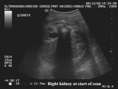
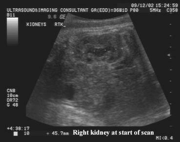
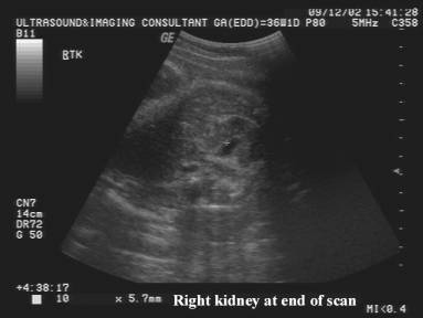
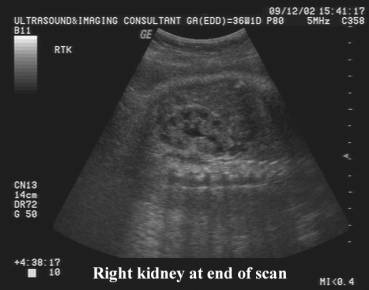
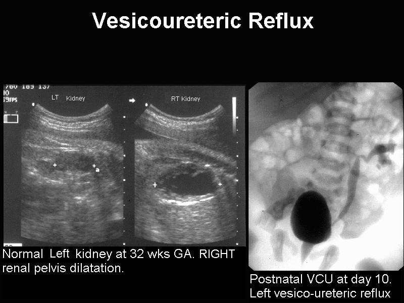
- Renal pelvic dilatation (most common sonographic finding).
- Ultrasound may not be able to distinguish vesicoureteric reflux from obstructive uropathy of the lower urinary tract in the presence of hydronephrosis, hydroureter and megacystis (4).
- Amniotic fluid volume is normal.
- Fetal urinoma may be present (2).
- The diagnosis may be made by vesico-infusion. A needle is placed into a distended fetal bladder after urine samples have been obtained. Saline is injected and the fetus examined for progressive dilatation of the renal pelvis as well as for spontaneous emptying of the fetal bladder thus excluding bladder outlet obstruction (2).
|
|
|
|
|
|
|
|
|
REFERENCES |
- Hiraoka M, Kasuga K, Hori C et.al. Ultrasonic indicators of ureteric reflux in the newborn. Lancet 1994;343:519-520.
- Quintero RA, Johnson MP, Arias F et.al. In utero sonographic diagnosis of vesicoureteral reflux by percutaneous vesicoinfusion. Ultrasound Obstet Gynecol 1995;6:386-389.
- Elder J. Commentary: importance of antenatal diagnosis of vesicoureteric reflux. J Urol 1992;148:1750.
- Reuter K, Lebowitz R. Massive vesicoureteral reflux mimicking posterior urethral valves in a fetus. J Clin Ultrasound 1985;13:584.