|
URETEROCELE |
A ureterocele is the
ballooning of the submucosal segment of the anterior
wall of the ureter into the bladder lumen.
It that may occur in single ureter
systems but when ectopic are usually associated with
a duplication anomaly. Bilateral ureteroceles
can occur
CLASSIFICATION |
- Simple (unassociated with a duplex collecting system).
- Complex (associated with a duplex collecting system).
- Intravesical ureterocele.
- Ectopic ureterocele (ureteroceles in children are usually ectopic with the orifice entering the urethra in 60% of cases).
Proposed ureterocele etiologies include ureteral meatal obstruction, incomplete muscularization of the distal ureter, and excessive dilatation of the ureter as it incorporates embryologically into the bladder.
ULTRASOUND |
- Usually unilateral (bilateral in 10-15% of cases) (1-5).
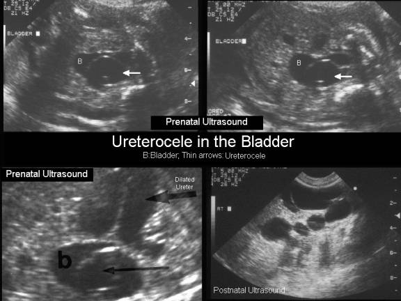
- Thin walled sonolucent mass predominantly within the bladder. If the bladder is partially filled a ureterocele may be missed.
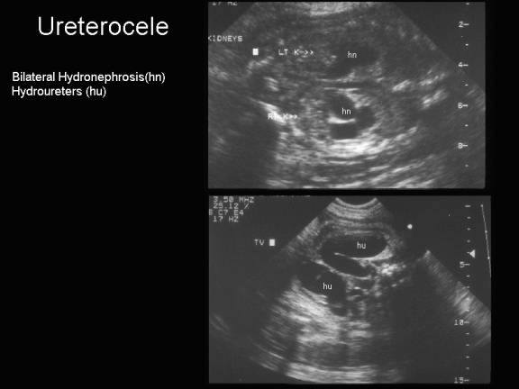
- Hydronephrosis and hydroureter.
- The majority involves a duplicated system draining the upper pole of a hydronephrotic or dysplastic kidney.
- If large enough it may prolapse into the bladder resulting in severe outflow obstruction.
- The renal parenchyma associated with a ureterocele is usually dysplastic and has little or no function.
- They have been reported to "vanish" (2).
- One report suggested they could simulate a pseudoseptated bladder (6).
- Occasional nonvisualization of a ureterocele in a bladder, particularly during fetal life may be the result of compression with change in intravesicle pressure.
- They may exist and be imaged outside the bladder simulating a fetus with such cystic masses as ovarian cyst or anterior meningocele (6,7).
|
|
|
|
|
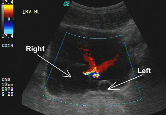
Postnatal
ultrasound of bilateral ureteroceles. Note the
bilateral ureteric jets suggesting that the ureteroceles are not causing any obstruction |
|
|
ASSOCIATED ANOMALIES |
- Usually isolated.
- Crossed renal ectopia.
- Abdominal testes.
- Cardiac anomalies.
COMPLICATIONS (2,3) |
- Obstruction of the ipsilateral ureter.
- Obstruction of the contralateral ureter or bladder outlet if it is large enough.
- Cystic dysplasia in the upper pole of the kidney.
REFERENCES |
- Garmel SH, Crombleholme TM, Cendron M et.al. The vanishing fetal ureterocele: a cause for concern. Prenat Diagn 1996;16:354-356.
- Bauer SB, Perlmutter AD, Retik AB.
Anomalies of the upper urinary tract. In: Walsh PC, Retik
AB, Stamey TA (eds).
- R. Yang and H.L. Cohen, Pyelectasis. In: H.L. Cohen and C.K. Sivit, Editors, Fetal
and Pediatric Ultrasound: A Casebook Approach,
- 81. A. Nussbaum, J. Dorst, R. Jeffs et al., Ectopic ureterocele: their varied sonographic manifestations. Radiology 159 (1986), pp. 227–235.
- 82. W. Sepulveda, C. Campana, E. Carstens et al., Prenatal sonographic diagnosis of bilateral ureteroceles: The pseudoseptated fetal bladder. J Ultrasound Med 22 (2003), pp. 841–844.